J Korean Soc Spine Surg.
2016 Jun;23(2):121-126. 10.4184/jkss.2016.23.2.121.
Extensive Intradural Epidermoid Cysts with Cauda Equina Syndrome in the Lumbosacral Spine: Case Report
- Affiliations
-
- 1Department of Orthopedic Surgery, CHA Bundang Medical Center, CHA University, Korea. shinde@cha.ac.kr
- KMID: 2328006
- DOI: http://doi.org/10.4184/jkss.2016.23.2.121
Abstract
- STUDY DESIGN: A case report.
OBJECTIVES
To report a rare case of extensive epidermoid cysts in the lumbosacral spine. SUMMARY OF LITERATURE REVIEW: The intradural epidermoid cyst with extensive involvement is rare, and previous reports have reported only extensive intramedullary epidermoid cysts.
MATERIALS AND METHODS
A 75-year-old male presented with progressive motor weakness of both extremities beginning 3 days prior. MRI showed extensive intradural extramedullary epidermoid cysts in the lumbosacral region. We performed total laminectomy from the L1 to the L5 level, and the cystic mass was removed.
RESULTS
We confirmed the epidermoid cyst on histopathologic examination.
CONCLUSIONS
Extensive extramedullary epidermoid cysts are difficult to remove completely. Attempting complete removal may result in neurological deficit. Therefore, when surgical intervention is planned, the poor postoperative prognosis should be taken into consideration.
MeSH Terms
Figure
-
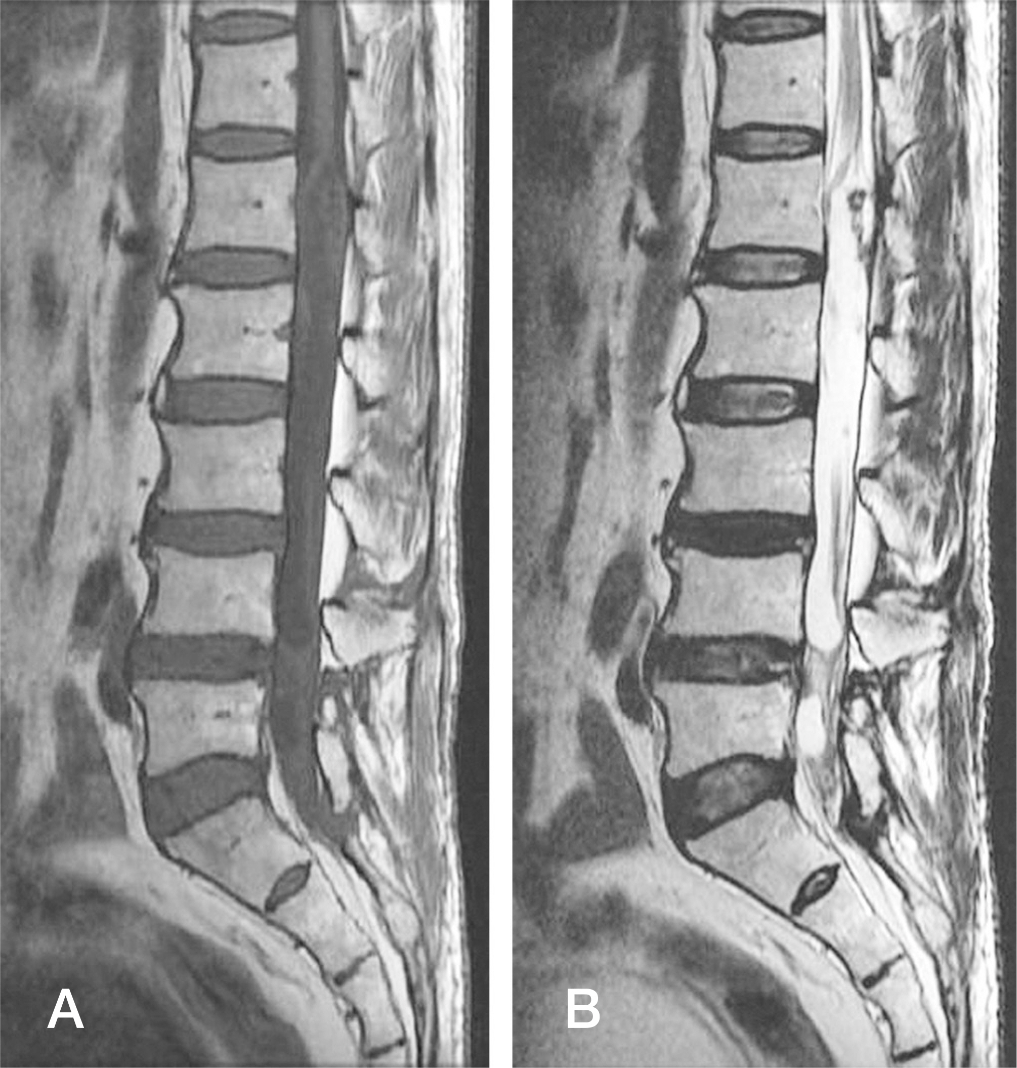
Fig. 1. Preoperative T1-weighted MRI sagittal (A) and T2-weighted MRI sagittal (B) images show an extramedullary elongated mass extending from the L1 to L5 level without widening of the spinal canal.
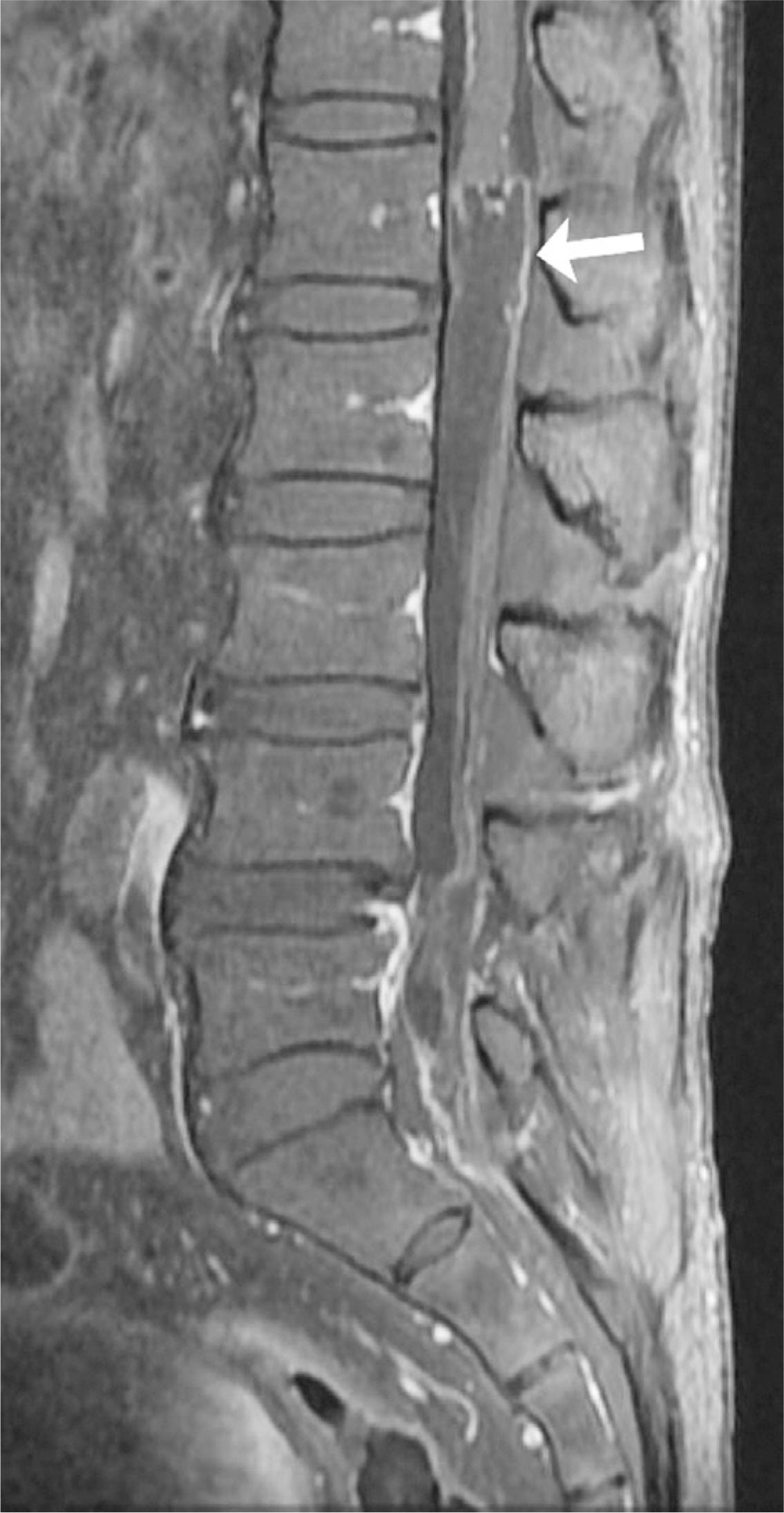
Fig. 2. Enhanced T1-weighted sagittal MRI reveals a thin rim enhancement around the mass.
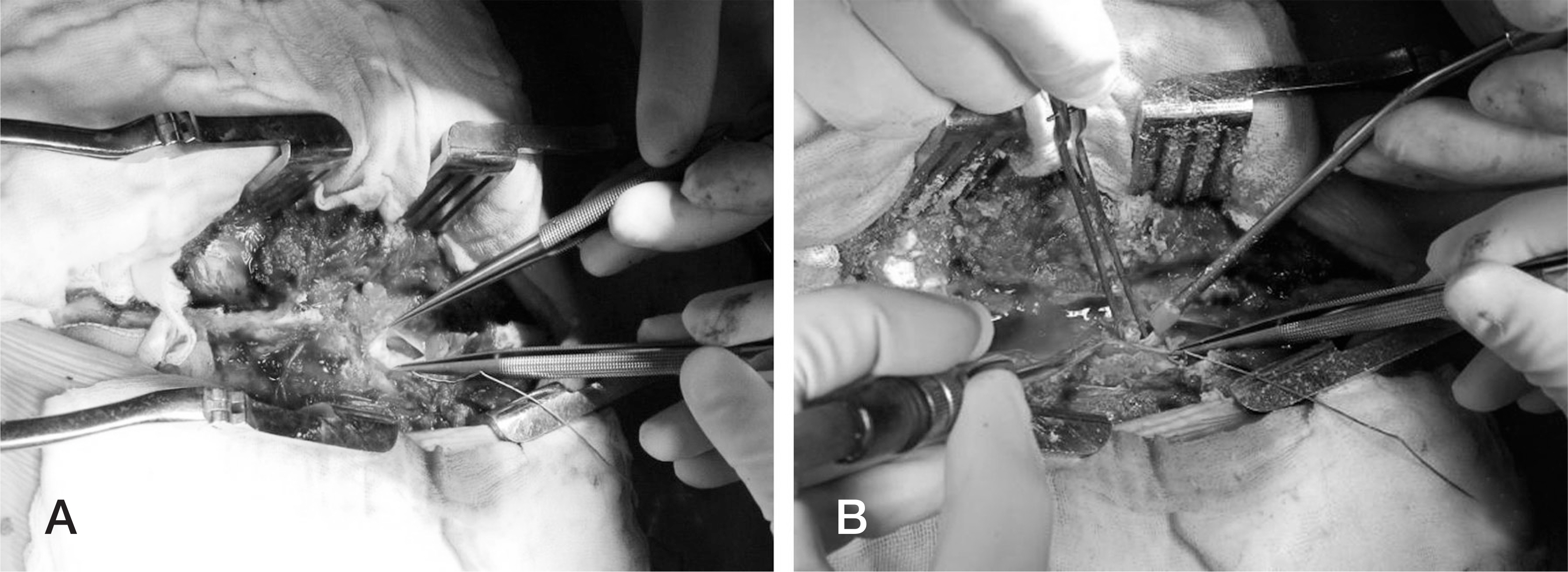
Fig. 3. (A-B) Intraoperative images show intradural epidermoid cyst.
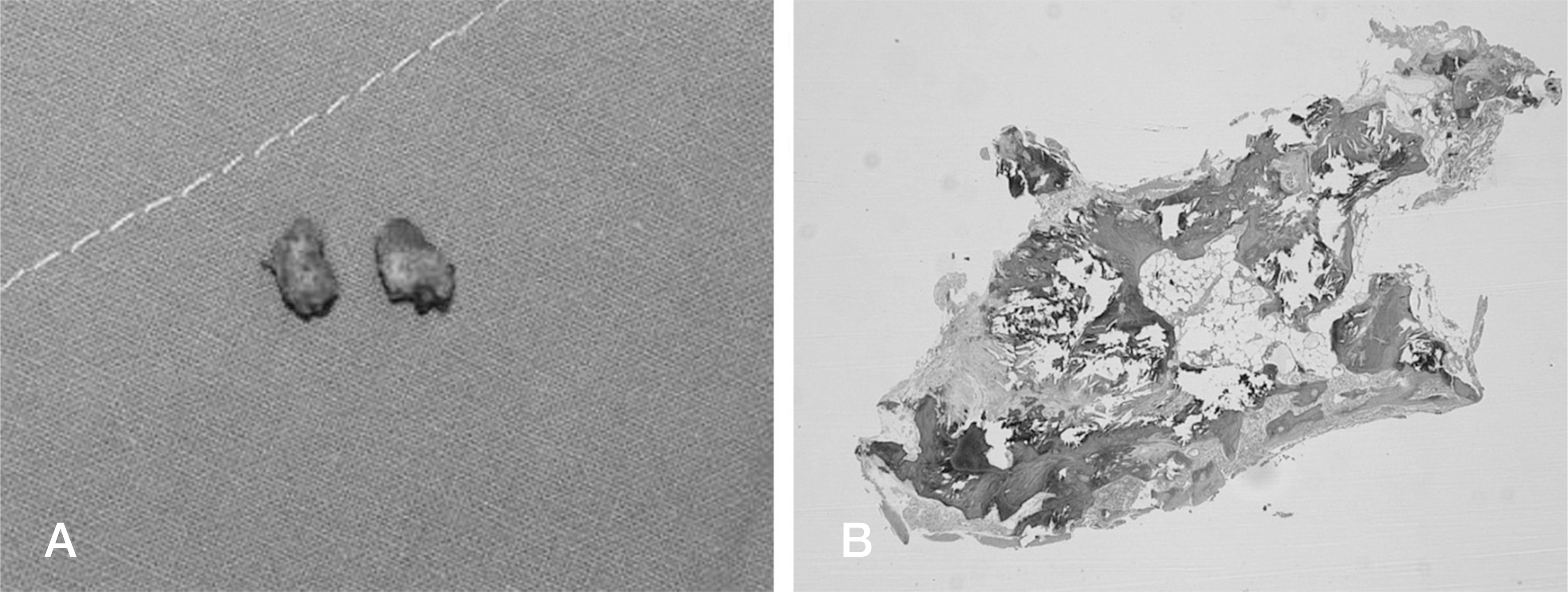
Fig. 4. Gross photo (A) and histopathological image (B, H&E stain, 1×) show a Calcified bone.

Fig. 5. A histopathological image (A, H&E stain, 1×) and another histopathological image (B, H&E stain, 4×) show an epidermoid cyst.
Reference
-
1. Teksam M, Casey SO, Michel E, et al. Intraspinalepi-dermoid cyst: diffusion weighted MRI. Neuroradiology. 2001; 43:572–4.2. Gardner DJ, O'Gorman AM, Blundell JE. Intraspinal epi-dermoid tumour: Late complication of lumbar puncture. CMAJ. 1989; 141:223–5.3. Cruveilhier J. Anatomic Pathologique du Corps Humain: Vol 1. Paris: Bailli6re, 1829, Book 2, Plate 6.4. Gonzalvo A, Hall N, McMahon JH, et al. Intramedullary spinal epidermoid cyst of the upper thoracic region. J Clin Neurosci. 2009; 16:142–4.
Article5. Ozaras N, Sariyildiz M, Demir S, et al. A neglected case admitted with paraplegia: An intradural extramedullary epidermoid cyst. J ClinExp Invest. 2012; 3:270.
Article6. Min Ho P, Tack Geun C, Jae Gon M, et al. Iatrogenic Intraspinal Epidermoid Cyst. Korean J Spine. 2014; 11:195–7.
Article7. Sarang G, Deepak R, Shrikant S, et al. Giantintradural in-tramedullary epidermoid cyst Report of two cases with var-ied presentations. Asian J Neurosurg. 2014; 9:244.8. Roux A, Mercier C, Larbrisseau A, et al. Intramedullary epidermoid cysts of the spinal cord: case report. J Neurosurg. 1992; 76:528–33.9. Alves AM, Norrell H. Intramedullary epidermoid tumors of the spinal cord. Report of a case and review of the literature. Int Surg. 1970; 54:239–43.10. Jee-Soo J, Sang-Ho L. Multiple Intramedullary and In-tradural Epidermoid Cystsin the Conus Medullaris and the Lumbar Spine. J Korean Neurosurg Soc. 2003; 33:512–3.11. Haber MD, Nguyen DD, Li S. Differentiation of Id-iopathic Spinal Cord Herniation from CSF-isointense IntraspinalExtramedullary Lesions Displacing the Cord. RadioGraphics. 2014; 34:313–9.
- Full Text Links
-
- Actions
-
Cited
- CITED
-
- Close
- Share
-
- Similar articles
-
- Intradural Epidermoid Cyst at Conus Medullaris and Cauda Equina of the Spine: A Case Report
- Cavernous Hemangioma of the Cauda Equina
- Huge Intradural Lumbar Disc Herniation Mimicking an Intradural Spinal Tumor: A Case Report
- Dorsal Herniation of Cauda Equina Due to Sequestrated Intradural Disc
- MRI of Cauda Equina Syndrome in Ankylosing Spondylitis: A Case Report
