MR Imaging Findings of Supratentorial Meningeal Hemangioblastoma: A Case Report
- Affiliations
-
- 1Department of Radiology, Jeju National University Hospital, Jeju, Korea. hoklee33@gmail.com
- 2Department of Pathology, Jeju National University Hospital, Jeju, Korea.
- KMID: 2327363
- DOI: http://doi.org/10.3348/jksr.2016.75.1.26
Abstract
- Hemangioblastomas account for 1.1-2.5% of intracranial neoplasms. These tumors most commonly occur in the cerebellum. A 77-year-old woman had a hemangioblastoma, which showed the supratentorial meningeal mass without any history of von Hippel-Lindau disease.
MeSH Terms
Figure
-
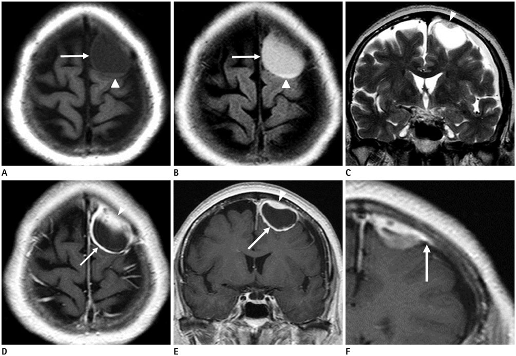
Fig. 1 A 77-year-old woman with supratentorial meningeal hemangioblastoma. A, B. Axial T1-weighted and T2 FLAIR image show a 3.7 cm, well-defined cystic mass (arrow) in the left frontal convexity. The cystic mass shows hypo signal intensity on T1-weighted MR image and high signal intensity on T2 FLAIR image. The cystic mass exhibits a high signal intensity portion (arrowheads) suggesting hemorrhage, on T1-weighted and T2 FLAIR images. C. Coronal T2-weighted image shows plaque-like mural nodule (arrowhead) of intermediate to high intensity signal. D, E. Axial and coronal gadolinium-enhanced T1-weighted fat-suppressed MR image show a rim-like enhancement of the mass (arrows) and plaque-like enhancing mural nodule (arrowheads). F. Coronal gadolinium-enhanced T1-weighted fat-suppressed MR image shows dural tail with adjacent plaque-like enhancing mural nodule (arrow).FLAIR = fluid attenuated inversion recovery
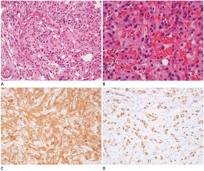
Fig. 2 Microphotographs showing numerous thin-walled vessels (A: H&E, × 200) and large, vacuolated stromal cells (B: H&E, × 400). Immunohistochemistry, highlighting the stromal cells, which are positive for NSE (C: × 200) and negative for EMA. Vascular endothelial cells reactive with CD31 (D: × 200) highlighting the positive reaction of stromal cells. Positive for NSE, negative for EMA, and positive for vascular endothelial cells of CD31 are inconsistent with meningioma. EMA = epithelial membrane antigen, H&E = hematoxylin and eosin, NSE = neuron specific enolase
Reference
-
1. Conway JE, Chou D, Clatterbuck RE, Brem H, Long DM, Rigamonti D. Hemangioblastomas of the central nervous system in Von Hippel-Lindau syndrome and sporadic disease. Neurosurgery. 2001; 48:55–62. discussion 62-63.2. Böhling T, Plate KH, Haltia MJ, Alitalo K, Neumann HP. World Health Organization classification of tumours pathology and genetics, tumours of the nervous system. Lyon: IARC Press;2000.3. Richard S, Graff J, Lindau J, Resche F. Von Hippel-Lindau disease. Lancet. 2004; 363:1231–1234.4. Peyre M, David P, Van Effenterre R, François P, Thys M, Emery E, et al. Natural history of supratentorial hemangioblastomas in von Hippel-Lindau disease. Neurosurgery. 2010; 67:577–587.5. Shin Y, Kim S, Lee HW, Bang H, Suh YL. Supratentorial hemangioblastoma with unusual features. Korean J Pathol. 2014; 48:462–465.6. Kim H, Park IS, Jo KW. Meningeal supratentorial hemangioblastoma in a patient with von hippel-lindau disease mimicking angioblastic menigioma. J Korean Neurosurg Soc. 2013; 54:415–419.7. Farrukh HM. Cerebellar hemangioblastoma presenting as secondary erythrocytosis and aspiration pneumonia. West J Med. 1996; 164:169–171.8. Bonneville F, Sarrazin JL, Marsot-Dupuch K, Iffenecker C, Cordoliani YS, Doyon D, et al. Unusual lesions of the cerebellopontine angle: a segmental approach. Radiographics. 2001; 21:419–438.9. Ho VB, Smirniotopoulos JG, Murphy FM, Rushing EJ. Radiologic-pathologic correlation: hemangioblastoma. AJNR Am J Neuroradiol. 1992; 13:1343–1352.10. Ono T, Sasajima T, Oda M, Mizoi K. [Cerebellar hemangioblastoma with marked pleomorphism: a case report]. No Shinkei Geka. 2012; 40:643–650.
- Full Text Links
-
- Actions
-
Cited
- CITED
-
- Close
- Share
-
- Similar articles
-
- Supratentorial Meningeal Hemangioblastoma: Case Report
- Meningeal Supratentorial Hemangioblastoma in a Patient with Von Hippel-Lindau Disease Mimicking Angioblastic Menigioma
- Congenital Cystic Supratentorial Hemangioblastoma Associated with Intracystic Hemorrhage: Case Report
- A Case of Supratentorial Hemangioblastoma Associated with Intraparenchymatous Hemorrhage: Case Report
- A Case of Hemangioblastoma in the Lateral Ventricle
