Ductal Carcinoma In Situ Arising in a Benign Phyllodes Tumor: A Case Report
- Affiliations
-
- 1Department of Radiology, Dongguk University Ilsan Hospital, Dongguk University College of Medicine, Goyang, Korea. dbkim@dumc.or.kr
- 2Department of Pathology, Dongguk University Ilsan Hospital, Dongguk University College of Medicine, Goyang, Korea.
- 3Department of Surgery, Dongguk University Ilsan Hospital, Dongguk University College of Medicine, Goyang, Korea.
Abstract
- A 42-year-old woman was presented with an ovoid mass detected on a mammography. Her physical examination revealed a 2 cm ill-defined mass in the right upper outer breast. A sonogram demonstrated a 1.9 cm ovoid, partially microlobulated and partially well-circumscribed, and an isoechoic mass with increased vascularity on Doppler imaging. Surgical excision was performed and the pathology revealed ductal carcinoma in situ (DCIS) in a phyllodes tumor. DCIS within a phyllodes tumor is a very rare event. Here, we report on a case of DCIS in a phyllodes tumor.
MeSH Terms
Figure
-
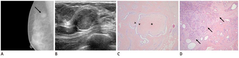
Fig. 1 A 42-year-old woman with DCIS in a phyllodes tumor. A. A mediolateral oblique mammogram shows a 2.2 × 1.4 cm sized, ovoid, circumscribed, and iso-dense mass in the upper portion of the right breast (arrow). B. A sonogram shows an ovoid, partially circumscribed, partially indistinct, and isoechoic mass in the right breast, 10 o'clock direction. Doppler US (not shown) showed vascularity in the mass. C. A well-defined tumor shows mildly cellular stroma (asterisk) and epithelial-lined cleft (arrowheads), exhibiting a leaf-like pattern. The stromal component revealed benign phyllodes tumor (Hematoxylin & Eosin staining, × 40). D. A 1.2 cm sized cribriform DCIS (arrows) is identified within the phyllodes tumor (Hematoxylin & Eosin staining, × 40). Note.-DCIS = ductal carcinoma in situ, US = ultrasound
Reference
-
1. Tavassoli FA, Devilee P. World Health Organization Classification of Tumours. Pathology and Genetics of Tumours of Breast and Female Genital Organs. 2003. Lyon: IARC Press;100–102.2. Rosen PP. Rosen's Breast Pathology. 2008. 3rd ed. Philadelphia: Lippincott Williams & Wilkins;724–729.3. Grove A, Deibjerg Kristensen L. Intraductal carcinoma within a phyllodes tumor of the breast: a case report. Tumori. 1986. 72:187–190.4. Deodhar KK, Baraniya JB, Naresh KN, Shinde SR, Chinoy RF. Cancerization of phyllodes tumour. Histopathology. 1997. 30:98–99.5. Nishimura R, Hasebe T, Imoto S, Mukai K. Malignant phyllodes tumour with a noninvasive ductal carcinoma component. Virchows Arch. 1998. 432:89–93.6. Lim SM, Tan PH. Ductal carcinoma in situ within phyllodes tumour: a rare occurrence. Pathology. 2005. 37:393–396.7. Nomura M, Inoue Y, Fujita S, Sakao J, Hirota M, Souda S, et al. A case of noninvasive ductal carcinoma arising in malignant phyllodes tumor. Breast Cancer. 2006. 13:89–94.8. Yamaguchi R, Tanaka M, Kishimoto Y, Ohkuma K, Ishida M, Kojiro M. Ductal carcinoma in situ arising in a benign phyllodes tumor: report of a case. Surg Today. 2008. 38:42–45.9. Parfitt JR, Armstrong C, O'malley F, Ross J, Tuck AB. In-situ and invasive carcinoma within a phyllodes tumor associated with lymph node metastases. World J Surg Oncol. 2004. 2:46.10. Korula A, Varghese J, Thomas M, Vyas F, Korula A. Malignant phyllodes tumour with intraductal and invasive carcinoma and lymph node metastasis. Singapore Med J. 2008. 49:e318–e321.
- Full Text Links
-
- Actions
-
Cited
- CITED
-
- Close
- Share
-
- Similar articles
-
- Multi-Focal Lobular Carcinoma In Situ Arising in Benign Phyllodes Tumor: A Case Report
- Invasive Ductal Carcinoma Arising in a Recurrent Malignant Phyllodes Tumor: A Case Report
- A Positive Hybrid (HMW-CK and E-Cadherin) Carcinoma in situ Arising in a Phyllodes Tumor of the Breast: A Case Report
- Borderline Phyllodes Tumor with an Incidental Invasive Tubular Carcinoma and Lobular Carcinoma In Situ Component: A Case Report
- Invasive Ductal Carcinoma Originating from a Borderline Phyllodes Tumor in a Young Female: A Case Report
