Imaging Findings of Mucinous Tubular and Spindle Cell Carcinoma of the Kidney: A Case Report
- Affiliations
-
- 1Department of Diagnostic Radiology, Soonchunhyang University Bucheon Hospital, Soonchunhyang University College of Medicine, Bucheon, Korea. rad1995@schmc.ac.kr
- 2Department of Urology, Soonchunhyang University Bucheon Hospital, Soonchunhyang University College of Medicine, Bucheon, Korea.
- 3Department of Pathology, Soonchunhyang University Bucheon Hospital, Soonchunhyang University College of Medicine, Bucheon, Korea.
Abstract
- Mucinous tubular and spindle cell carcinoma of the kidney has been recognized as a distinct entity in the 2004 World Health Organization classification of adult renal tumors; it constitutes less than 1% of all the renal neoplasm. Radiological features of mucinous tubular and spindle cell carcinoma have been published in a small number of cases. This case report presents a case of mucinous tubular and spindle cell carcinoma, including CT and MR finding.
MeSH Terms
Figure
-
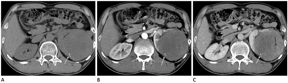
Fig. 1 CT finding of mucinous tubular and spindle cell carcinoma of the kidney. A. Precontrast CT scan of 54-year-old male shows isodense mass in upper pole of left kidney with focal calcification (white arrow). B, C. Contrast enhanced corticomedullary and nephrogenic phase CT scan of axial images show heterogeneous enhancement (black arrow) of well defined solid huge mass and focal calcification (white arrow).
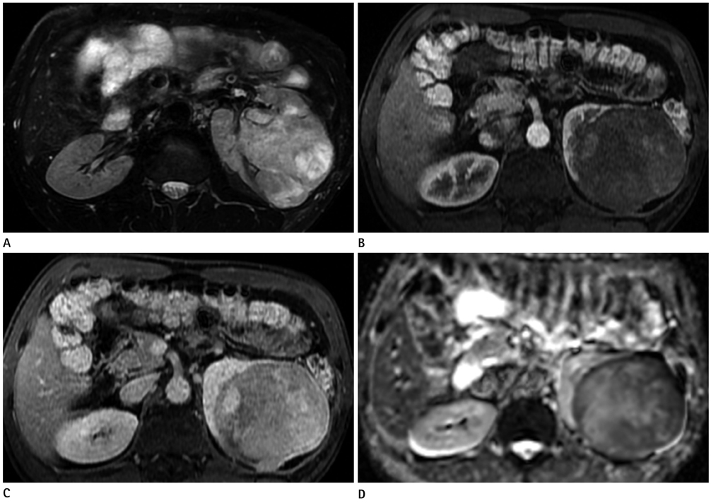
Fig. 2 MR finding of mucinous tubular and spindle cell carcinoma of the kidney. A. Axial T2-weighted fast spine echo MR image demonstrated a well marginated heterogeneously high signal intensity tumor in upper pole of left kidney. B, C. Gadolinium enhanced dynamic scan shows heterogeneous and delayed mild enhancement of tumor (30 seconds and 90 seconds, respectively). D. On diffusion weighted image (b value = 800), some area shows high signal intensity (not shown) and the corresponding apparent diffusion coefficient mapping MR image demonstrates diffusion restriction.
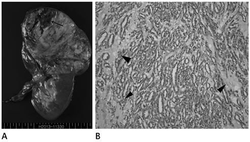
Fig. 3 Gross and microscopic finding of mucinous tubular and spindle cell carcinoma of the kidney. A. Specimen shows 13 cm sized well demarcated solid mass in upper pole of left kidney confined to renal capsule. The cut surface of tumor demonstrates yellow-gray color with hemorrhagic foci (balck arrow). B. On microscopic findings, tubular structures with spindle cells and mucinous component (arrowheads) are manifested (H&E, × 100).
Reference
-
1. Eble JN, Sauter G, Epstein JI, Sesterhenn IA. World Health Organization Classification of Tumours. Pathology and Genetics of Tumours of the Urinary System and Male Genital Organs. Lyon: IARC Press;2004.2. Parwani AV, Husain AN, Epstein JI, Beckwith JB, Argani P. Low-grade myxoid renal epithelial neoplasms with distal nephron differentiation. Hum Pathol. 2001; 32:506–512.3. Srigley J, Kapusta L, Reuter V, Amin M, Grignon D, Eble J, et al. Phenotypic, molecular and ultrastructural studies of a novel low grade renal epithelial neoplasm possibly related to the loop of Henle. Mod Pathol. 2002; 15:182A.4. Shanbhogue AK, Vikram R, Paspulati RM, MacLennan G, Verma S, Sandrasegaran K, et al. Rare (<1%) histological subtypes of renal cell carcinoma: an update. Abdom Imaging. 2012; 37:861–872.5. Sahni VA, Hirsch MS, Sadow CA, Silverman SG. Mucinous tubular and spindle cell carcinoma of the kidney: imaging features. Cancer Imaging. 2012; 12:66–71.6. Lima MS, Barros-Silva GE, Pereira RA, Ravinal RC, Tucci S Jr, Costa RS, et al. The imaging and pathological features of a mucinous tubular and spindle cell carcinoma of the kidney: a case report. World J Surg Oncol. 2013; 11:34.7. Tirumani SH, Assiri YI, Brimo F, Tsatoumas M, Reinhold C. Diffusion-weighted MR imaging of mucin-rich mucinous tubular and spindle cell carcinoma of the kidney: a case report. Clin Imaging. 2013; 37:775–777.8. Kim JK, Kim TK, Ahn HJ, Kim CS, Kim KR, Cho KS. Differentiation of subtypes of renal cell carcinoma on helical CT scans. AJR Am J Roentgenol. 2002; 178:1499–1506.9. Oliva MR, Glickman JN, Zou KH, Teo SY, Mortelé KJ, Rocha MS, et al. Renal cell carcinoma: t1 and t2 signal intensity characteristics of papillary and clear cell types correlated with pathology. AJR Am J Roentgenol. 2009; 192:1524–1530.10. Rosenkrantz AB, Hindman N, Fitzgerald EF, Niver BE, Melamed J, Babb JS. MRI features of renal oncocytoma and chromophobe renal cell carcinoma. AJR Am J Roentgenol. 2010; 195:W421–W427.
- Full Text Links
-
- Actions
-
Cited
- CITED
-
- Close
- Share
-
- Similar articles
-
- Mucinous Tubular and Spindle Cell Carcinoma of Kidney Occurring in a Patient with Pulmonary Adenocarcinoma
- Mucinous Tubular and Spindle Cell Carcinoma of the Kidney: Touch Imprint Cytologic and Histologic Findings: A Case Report
- Mucinous Tubular and Spindle Cell Carcinoma of the Kidney with Aggressive Behavior: An Unusual Renal Epithelial Neoplasm: A Case Report
- Tubular adenoma of the gallbladder with spindle cell metaplasia
- CT Findings of Mucinous Adenocarcinoma Arising from the Renal Calyx in Horseshoe Kidney: A Case Report
