J Korean Acad Oral Health.
2014 Jun;38(2):105-110.
In vitro quantification of occlusal caries lesion using QLF-D, ICDAS, and DIAGNOdent
- Affiliations
-
- 1Department of Preventive and Community Dentistry, Pusan National University School of Dentistry, Pusan National University, Yangsan, Korea. jsh0917@pusan.ac.kr
- 2Institute of Translational Dental Sciences, Pusan National University, Yangsan, Korea.
- 3Department of Preventive Dentistry & Public Oral Health, Yonsei University College of Dentistry, Seoul, Korea.
Abstract
OBJECTIVES
To compare the QLF-D method and the ICDAS and DIAGNOdent techniques for in vitro quantification of occlusal caries and to assess the histological features of the caries.
METHODS
One hundred and twenty-two extracted permanent teeth were selected, and the site of interest on the occlusal surface was examined using each detection method. The occlusal sites were classified according to the ICDAS II criteria based on the decision taken by two investigators, who have taken the ICDAS E-learning course. The examined site was then measured using the DIAGNOdent, and the peak value was recorded. In addition, by using the QLF-D, the occlusal site was photographed to obtain the DeltaFmax value. After all assessments were performed, the occlusal sites were vertically sectioned in order to assess the histological features. This was considered the gold standard. The histological criteria were graded using a 4-point scale as follows: S=sound (n=21), E1=limited enamel caries (n=27), E2=caries extending to the dento-enamel junction (n=49), D=caries involving the dentine (n=25).
RESULTS
An ICDAS code between 0 and 4 was assigned to all the occlusal sites, and this revealed the QLF-D value, which was between -95 to 0. The DIAGNOdent value was between 8 and 99. The correlation values of QLF-D, ICDAS, and DIAGNOdent with the histological features were 0.68, 0.58, and 0.46, respectively (P<0.01). A highly significant correlation was observed between QLF-D and the gold standard, which showed a moderate correlation and an acceptable correlation was observed with ICDAS (r=0.75, P<0.01). A statistically significant difference was observed in the average QLF-D values of each histological grade i.e., -28.5 (S), -53.7 (E1), -68.1 (E2), and -84.4 (D).
CONCLUSIONS
The QLF-D showed a significant correlation with the ICDAS and histological features. Therefore, visual inspection with QLF-D would improve the detection accuracy and ensure early diagnosis of dental caries.
Keyword
Figure
-
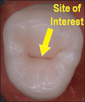
Fig. 1. Site of interest of each occulsal surface. the site was selected by one examiner.

Fig. 2. An example of lesion analysis procedure using QLF-D.

Fig. 3. ICDAS and histological criteria used in this study.
Reference
-
References
1. Pitts N, Stamm J. International Consensus Workshop on Caries Clinical Trials (ICW-CCT)-final consensus statements: agreeing where the evidence leads. J Dent Res. 2004; 83(suppl 1):C125–C128.
Article2. World Health Organization. Oral health surveys-basic method. 4th ed. Geneva. 1997.3. Ministry of Health and Welfare. 2010 Korean National Oral Health Survey: Ⅰ. Survey Process Report. Seoul: Ministry of Health & Welfare;2011. p. 71–72.4. Ismail A, Sohn W, Tellez M, Amaya A, Sen A, Hasson H, et al. The International Caries Detection and Assessment System (ICDAS): an integrated system for measuring dental caries. Community Dent Oral Epidemiol. 2007; 35:170–178.
Article5. Kim BI. QLF Concept and Clinical Implementation. Journal Kor Dent Ass. 2011; 49:443–450.6. van der Veen MH, de Josselin de Jong E. Application of quantitative light-induced fluorescence for assessing early caries lesions. Monogr Oral Sci. 2000; 17:144–162.
Article7. Pretty IA. Caries detection and diagnosis: novel technologies. J Dent. 2006; 34:727–739.
Article8. Lee BJ. Use of laser fluorescence device ‘DIAGNODent’ for detecting caries. Journal Kor Dent Ass. 2011; 49:461–471.9. Pinelli C, Campos Serra M, de Castro Monteiro Loffredo L. Validity and reproducibility of a laser fluorescence system for detecting the activity of white-spot lesions on free smooth surfaces in vivo. Caries Res. 2002; 36:19–24.
Article10. Lussi A, Hibst R, Paulus R. DIAGNOdent: an optical method for caries detection. J Dent Res. 2004; 83(suppl 1):C80–C83.
Article11. Farah R, Drummond B, Swain M, Williams S. Relationship between laser fluorescence and enamel hypomineralisation. J Dent. 2008; 36:915–921.
Article12. Bader JD, Shugars DA. A systematic review of the performance of a laser fluorescence device for detecting caries. J Am Dent Assoc. 2004; 135:1413–1426.
Article13. Fejerskov O, Nyvad B, Kidd E.A.M.Pathology of dental caries. Fejerskov O, editor. Dental caries: the disease and its clinical man-agemen. 2nd ed.Oxford: Backwell Munksgaad Ltd.;2008. p. 19–48.14. Lussi A. Validity of diagnostic and treatment decisions of fissure caries. Caries Res. 1991; 25:296–303.
Article15. Weerheijm K, Van Amerongen W, Eggink C. The clinical diagnosis of occlusal caries: a problem. ASDC J Dent Child. 1988; 56:196–200.16. Ekstrand K, Ricketts D, Kidd E. Reproducibility and accuracy of three methods for assessment of demineralization depth on the occlusal surface: an in vitro examination. Caries Res. 1997; 31:224–231.
Article17. Stookey GK. Quantitative light fluorescence: a technology for early monitoring of the caries process. Dent Clin North Am. 2005; 49:753–770.
Article18. Gomez J, Zakian C, Salsone S, Pinto SC, Taylor A, Pretty IA, et al. In vitro performance of different methods in detecting occlusal caries lesions. J Dent. 2013; 41:180–186.
Article19. Bamzahim M, Shi X-Q, Angmar-Månsson B. Occlusal caries detection and quantification by DIAGNOdent and Electronic Caries Monitor: in vitro comparison. Acta Odontologica. 2002; 60:360–364.
Article20. Lussi A, Megert B, Longbottom C, Reich E, Francescut P. Clinical performance of a laser fluorescence device for detection of occlusal caries lesions. Eur J Oral Sci. 2001; 109:14–19.
Article21. Kavo Dental GmbH. User Instructions DIAGNOdent pen. 2009.22. Ferreira Zandoná A, Santiago E, Eckert G, Fontana M, Ando M, Zero D. Use of ICDAS combined with quantitative light-induced fluorescence as a caries detection method. Caries Res. 2010; 44:317–322.
Article23. Diniz MB, Rodrigues JA, Hug I, De Cássia Loiola Cordeiro R, Lus-si A. Reproducibility and accuracy of the ICDAS-II for occlusal caries detection. Community Dent Oral Epidemiol. 2009; 37:399–404.
Article
- Full Text Links
-
- Actions
-
Cited
- CITED
-
- Close
- Share
-
- Similar articles
-
- Detection of proximal caries using quantitative light-induced fluorescence-digital and laser fluorescence: a comparative study
- Assessment of the Caries Detection Ability of Quantitative Light-induced Fluorescence (QLF) in Primary Teeth
in vitro - Evaluation of Detection Ability of a Quantitative Light-Induced Fluorescence Digital Device for Initial Secondary Caries Lesion
- Detecting of Proximal Caries in Primary Molars using Pen-type QLF Device
- Detection of early changes in caries lesion using QLF-D and OCT
