Tuberc Respir Dis.
2013 Nov;75(5):222-224.
A Case of Bilateral Giant Bullae in Young Adult
- Affiliations
-
- 1Division of Pulmonary and Critical Care Medicine, Department of Internal Medicine and Lung Institute, Seoul National University College of Medicine, Seoul, Korea.
- 2Department of Thoracic Surgery, Seoul Metropolitan Government Seoul National University Boramae Medical Center, Seoul National University College of Medicine, Seoul, Korea.
- 3Division of Pulmonary and Critical Care Medicine, Department of Internal Medicine, Seoul Metropolitan Government Seoul National University Boramae Medical Center, Seoul National University College of Medicine, Seoul, Korea. kimdkmd@snu.ac.kr
Abstract
- Giant bullae are large bullae occupying at least one-third of the hemithorax and surgical bullectomy is the treatment of choice. We report a case with symptomatic giant bullae which were resected successfully. A 35-year-old man presented with bilateral giant bullae that occupied almost the entire left hemithorax and a third of the right hemithorax. He was a current smoker with a 30 pack-year history and he presented with dyspnea on exertion. An elective surgical bullectomy was performed with video-assisted thoracoscopic surgery. The patient recovered without any adverse events and stayed well for 1 month after surgery.
Keyword
Figure
-
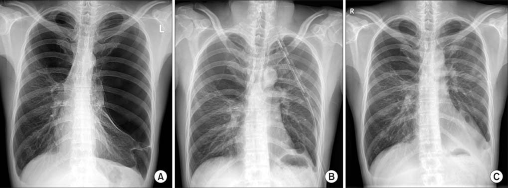
Figure 1 Representative chest radiographies. (A) Bilateral giant bullae occupying almost the entire left hemithorax and a third of the right thorax; preoperative period. (B) Postoperative period. (C) Follow-up 1 month after surgery.
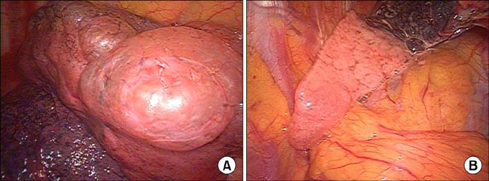
Figure 2 Intraoperative findings. (A) Giant bulla at the left upper lobe apex. (B) Atelectasis left lower lobe.
Reference
-
1. Burke RM. Vanishing lungs: a case report of bullous emphysema. Radiology. 1937; 28:367–371.2. Palla A, Desideri M, Rossi G, Bardi G, Mazzantini D, Mussi A, et al. Elective surgery for giant bullous emphysema: a 5-year clinical and functional follow-up. Chest. 2005; 128:2043–2050.3. Stern EJ, Webb WR, Weinacker A, Muller NL. Idiopathic giant bullous emphysema (vanishing lung syndrome): imaging findings in nine patients. AJR Am J Roentgenol. 1994; 162:279–282.4. Kayawake H, Chen F, Date H. Surgical resection of a giant emphysematous bulla occupying the entire hemithorax. Eur J Cardiothorac Surg. 2013; 43:e136–e138.5. Bradshaw DA, Murray KM, Amundson DE. Spontaneous regression of a giant pulmonary bulla. Thorax. 1996; 51:549–550.6. Roberts L, Putman CE, Chen JT, Goodman LR, Ravin CE. Vanishing lung syndrome: upper lobe bullous pneumopathy. Rev Interam Radiol. 1987; 12:249–255.7. Sharma N, Justaniah AM, Kanne JP, Gurney JW, Mohammed TL. Vanishing lung syndrome (giant bullous emphysema): CT findings in 7 patients and a literature review. J Thorac Imaging. 2009; 24:227–230.8. Waseem M, Jones J, Brutus S, Munyak J, Kapoor R, Gernsheimer J. Giant bulla mimicking pneumothorax. J Emerg Med. 2005; 29:155–158.
- Full Text Links
-
- Actions
-
Cited
- CITED
-
- Close
- Share
-
- Similar articles
-
- Expeditious Resolution of Giant Bullae with Endobronchial Valves and Percutaneous Catheter Insertion
- A Case of Giant Cell Tumor in the Lumbar Vertebra
- Unilateral Giant Bullae: Pulmonary Placental Transmogrification Should Be Kept in Mind: Case Reports
- A Case Report of Giant Hydronephrosis
- Outcomes of Contralateral Bullae in Primary Spontaneous Pneumothorax
