A Case of Pulmonary Lymphangiomatosis
- Affiliations
-
- 1Department of Pulmonary and Critical Care Medicine, Asan Medical Center, University of Ulsan College of Medicine, Seoul, Korea. skysong3@hanmail.net
Abstract
- Pulmonary lymphangiomatosis is a rare disorder involving the entire intrathoracic lymphatic system from the mediastinum to the pleura. Pulmonary lymphangiomatosis mostly occurs in children and young adults without gender predilection. Although it is pathologically benign, it shows a progressive and fatal course with variable initial presentation. We now report a case of pulmonary lymphangiomatosis in a 35-year-old man. He presented with hemoptysis 6 months previously. Chest x-ray and a chest computed tomography scan showed diffuse interstitial thickening with left pleural effusion. Chylothorax was confirmed by thoracentesis. Lymphangiography showed dilated and tortuous lymphatic channels. Surgical lung biopsy revealed proliferation of complex anastomosing lymphatic channels. He was diagnosed with pulmonary lymophangiomatosis. Closed thoracostomy and chemical pleurodesis were done and the dyspnea was reduced.
MeSH Terms
Figure
-
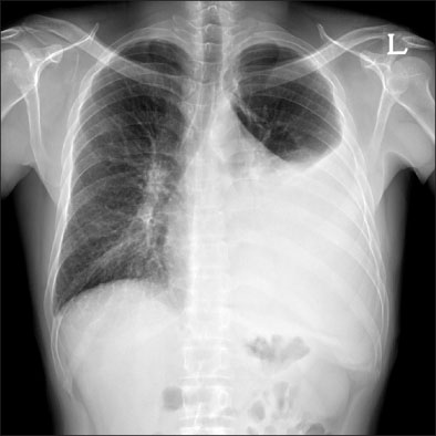
Figure 1 Chest X-ray shows peripheral and axial interstitial thickening in both lungs with left pleural effusion.
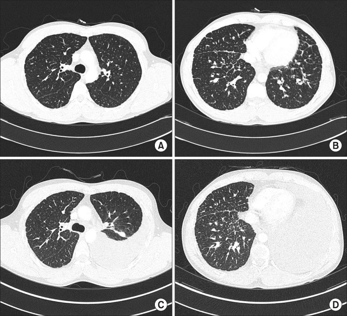
Figure 2 (A, B) Chest CT shows peripheral and axial interstitial thickening in both lungs (1st admission). (C, D) Chest CT shows newly appeared large amount of pleural effusion and passive atelectasis in left lung. Peripheral and axial interstitial thickening in both lungs shows no significant interval changes since 1st admission (2nd admission).
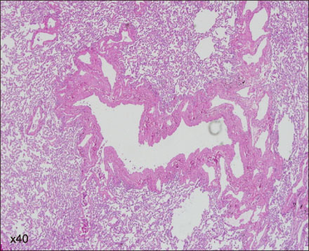
Figure 3 Surgical lung biopsy shows proliferation of complex anastomosing lymphatic channels in the interstitium (H&E stain, ×40).
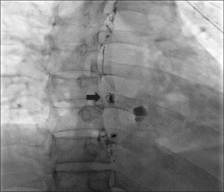
Figure 4 Lymphangiogram shows tortuous lymphatic channels and lymphatic leakage (arrow) idenitified at the medial aspect of the descending thoracic aorta.

Figure 5 Chest CT after lymphangiography shows multifocal areas of residual contrast material within abnormal lymphatic spaces in bilateral lower mediastinum and left lower lobe. CT: computed tomography.
Reference
-
1. Faul JL, Berry GJ, Colby TV, Ruoss SJ, Walter MB, Rosen GD, et al. Thoracic lymphangiomas, lymphangiectasis, lymphangiomatosis, and lymphatic dysplasia syndrome. Am J Respir Crit Care Med. 2000. 161:1037–1046.2. Hilliard RI, McKendry JB, Phillips MJ. Congenital abnormalities of the lymphatic system: a new clinical classification. Pediatrics. 1990. 86:988–994.3. Tazelaar HD, Kerr D, Yousem SA, Saldana MJ, Langston C, Colby TV. Diffuse pulmonary lymphangiomatosis. Hum Pathol. 1993. 24:1313–1322.4. Raman SP, Pipavath SN, Raghu G, Schmidt RA, Godwin JD. Imaging of thoracic lymphatic diseases. AJR Am J Roentgenol. 2009. 193:1504–1513.5. Swensen SJ, Hartman TE, Mayo JR, Colby TV, Tazelaar HD, Müller NL. Diffuse pulmonary lymphangiomatosis: CT findings. J Comput Assist Tomogr. 1995. 19:348–352.6. Yekeler E, Dursun M, Yildirim A, Tunaci M. Diffuse pulmonary lymphangiomatosis: imaging findings. Diagn Interv Radiol. 2005. 11:31–34.7. Jang HJ, Lee KS, Han J. Intravascular lymphomatosis of the lung: radiologic findings. J Comput Assist Tomogr. 1998. 22:427–429.8. Molitch HI, Unger EC, Witte CL, vanSonnenberg E. Percutaneous sclerotherapy of lymphangiomas. Radiology. 1995. 194:343–347.9. Rostom AY. Treatment of thoracic lymphangiomatosis. Arch Dis Child. 2000. 83:138–139.10. Reinhardt MA, Nelson SC, Sencer SF, Bostrom BC, Kurachek SC, Nesbit ME. Treatment of childhood lymphangiomas with interferon-alpha. J Pediatr Hematol Oncol. 1997. 19:232–236.11. Kandil A, Rostom AY, Mourad WA, Khafaga Y, Gershuny AR, el-Hosseiny G. Successful control of extensive thoracic lymphangiomatosis by irradiation. Clin Oncol (R Coll Radiol). 1997. 9:407–411.12. Valentine VG, Raffin TA. The management of chylothorax. Chest. 1992. 102:586–591.13. Calabrese PR, Frank HD, Taubin HL. Lymphangiomyomatosis with chylous ascites: treatment with dietary fat restriction and medium chain triglycerides. Cancer. 1977. 40:895–897.14. Kransdorf MJ. Benign soft-tissue tumors in a large referral population: distribution of specific diagnoses by age, sex, and location. AJR Am J Roentgenol. 1995. 164:395–402.15. Cho YS, Yoo JH, Son SY, Cho HL, Kim SJ, Han MS, et al. Pulmonary lymphangiomatosis. Tuberc Respir Dis. 1999. 47:533–537.
