Tuberc Respir Dis.
2008 Dec;65(6):457-463.
Correlation between Expiratory Increase of Lung Attenuation and Age and Smoking in the Subjects with Normal Inspiratory Low Dose CT and Pulmonary Function Test
- Affiliations
-
- 1Respiratory Center, Seoul National University Bundang Hospital, Seongnam, Korea. lkwrad@radiol.snu.ac.kr
- 2Department of Internal Medicine, Seoul National University Bundang Hospital, Seongnam, Korea.
- 3Department of Radiology, Seoul National University Bundang Hospital, Seongnam, Korea.
- 4Department of Internal Medicine, Hallym University Sacred Heart Hospital, Seoul, Korea.
Abstract
-
BACKGROUND: The attenuation of the lung parenchyma increases on expiration as a consequence of decreased air in the lung. Expiratory CT scans have been used to show air trapping in patients with chronic airway disease and diffuse parenchymal disease and also in asymptomatic smokers. Although there have been several reports investigating the regional air trapping on a expiratory CT scan, there have been only a few reports evaluating the changes of whole lung attenuation with considering its clinical significance, and especially in healthy subjects. The purpose of this study was to evaluate the correlation of an expiratory increase of lung attenuation with age and smoking in healthy subjects.
METHODS
Asymptomatic subjects who underwent a low dose chest CT scan as part of a routine check-up and who showed normal spirometry and a normal inspiratory CT scan were recruited for this study. We excluded the subjects with significant regional air trapping seen on their expiratory CT scan. Lung attenuation was measured at 24 points of both the inspiratory and expiratory CT scans, respectively, for 100 subjects. The correlations between an expiratory increase of the lung attenuation and the amount of smoking, the patient's age and the results of spirometric test were assessed.
RESULTS
There were 87 men and 13 women included in this study. Their median age was 49.0 years old (range: 25~71). Sixty current smokers, 24 ex-smokers and 16 non-smokers were included. As age increased, the expiratory increase of lung attenuation was reduced at every measuring points (r=-0.297~-0.487, Pearson correlation). The statistical significance was maintained after controlling for the effect of smoking. Smoking was associated with a reduction of the expiratory increase of lung attenuation. But the significance was reduced after controlling for the patient's age. The FEV1, FVC, FEV1/FVC and FEF25~75% were not associated with an expiratory increase of lung attenuation.
CONCLUSION
The expiratory increase of lung attenuation in subjects with a normal inspiratory CT scan was negatively correlated with age. It was also reduced in heavy smokers. It may reflect aging and the smoking related changes.
Keyword
Figure
-
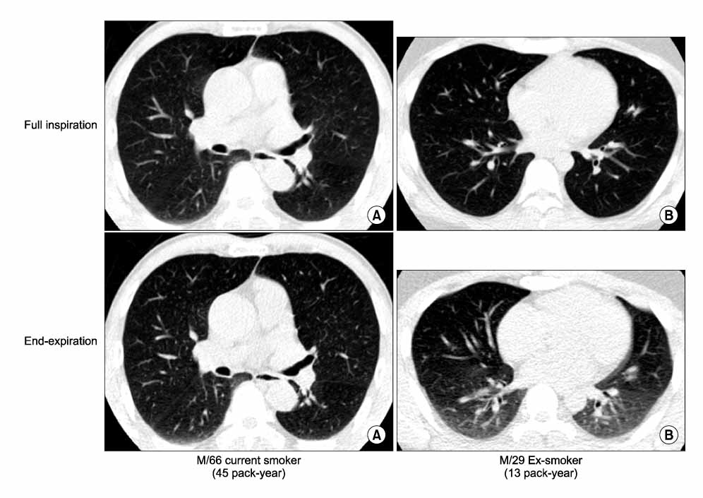
Figure 1 Examples of expiratory CT findings. (A) Paired inspiratory and expiratory CT scan of 66 years old current smoking male with 45 pack-year of smoking amount. Expiratory CT scan shows minimal change of lung density (bottom). (B) Paired inspiratory and expiratory CT scan of 29 years old ex-smoking male with 13 pack-year of smoking amount. Expiratory CT scan shows increased lung density compared with inspiratory CT scan (bottom).
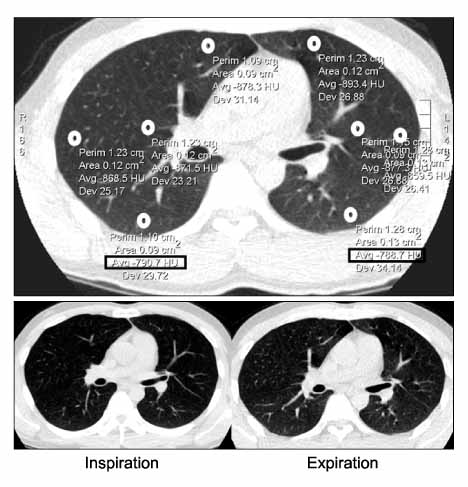
Figure 2 Example of lung attenuation measurement.
Reference
-
1. Naidich DP, Webb WR, Grenier PA, Harkin TJ, Gefter WB. Functinal imaging of the airways. Imaging of the airways. Functional and Radiologic correlation. 2005. 1st ed. Philadelphia: Lippincott Williams & Wilkins;177–211.2. Hansell DM. Small airway diseases: detection and insights with computed tomography. Eur Respir J. 2001. 17:1294–1313.3. Kazerooni EA. High-resolution CT of the lungs. AJR Am J Roentgenol. 2001. 177:501–519.4. Grenier PA, Beigelman-Aubry C, Fetita C, Preteux F, Brauner MW, Lenoir S. New frontiers in CT imaging of airway disease. Eur Radiol. 2002. 12:1022–1044.5. Austin JH, Muller NL, Friedman PJ, Hansell DM, Naidich DP, Remy-Jardin M, et al. Glossary of terms for CT of the lungs: recommendations of the Nomenclature Committee of the Fleischner Society. Radiology. 1996. 200:327–331.6. Arakawa H, Webb WR, McCowin M, Katsou G, Lee KN, Seitz RF. Inhomogeneous lung attenuation at thin-section CT: diagnostic value of expiratory scans. Radiology. 1998. 206:89–94.7. de Jong PA, Muller NL, Pare PD, Coxson HO. Computed tomographic imaging of the airways: relationship to structure and function. Eur Respir J. 2005. 26:140–152.8. Lucidarme O, Coche E, Cluzel P, Mourey-Gerosa I, Howarth N, Grenier P. Expiratory CT scans for chronic airway disease: correlation with pulmonary function test results. AJR Am J Roentgenol. 1998. 170:301–307.9. Terasaki H, Fujimoto K, Muller NL, Sadohara J, Uchida M, Koga T, et al. Pulmonary sarcoidosis: comparison of findings of inspiratory and expiratory high-resolution CT and pulmonary function tests between smokers and nonsmokers. AJR Am J Roentgenol. 2005. 185:333–338.10. Arakawa H, Niimi H, Kurihara Y, Nakajima Y, Webb WR. Expiratory high-resolution CT: diagnostic value in diffuse lung diseases. AJR Am J Roentgenol. 2000. 175:1537–1543.11. Spaggiari E, Zompatori M, Verduri A, Chetta A, Bna C, Ormitti F, et al. Early smoking-induced lung lesions in asymptomatic subjects: correlations between high resolution dynamic CT and pulmonary function testing. Radiol Med. 2005. 109:27–39.12. Verschakelen JA, Scheinbaum K, Bogaert J, Demedts M, Lacquet LL, Baert AL. Expiratory CT in cigarette smokers: correlation between areas of decreased lung attenuation, pulmonary function tests and smoking history. Eur Radiol. 1998. 8:1391–1399.13. Mastora I, Remy-Jardin M, Sobaszek A, Boulenguez C, Remy J, Edme JL. Thin-section CT finding in 250 volunteers: assessment of the relationship of CT findings with smoking history and pulmonary function test results. Radiology. 2001. 218:695–702.14. Lee KW, Chung SY, Yang I, Lee Y, Ko EY, Park MJ. Correlation of aging and smoking with air trapping at thin-section CT of the lung in asymptomatic subjects. Radiology. 2000. 214:831–836.15. Webb WR, Muller NL, Naidich DP. Normal lung anatomy. High-Resolution CT of the Lung. 2001. 3rd ed. Philadelphia: Lippincott Williams & Wilkins;49–69.16. Kauczor HU, Hast J, Heussel CP, Schlegel J, Mildenberger P, Thelen M. CT attenuation of paired HRCT scans obtained at full inspiratory/expiratory position: comparison with pulmonary function tests. Eur Radiol. 2002. 12:2757–2763.17. Webb WR, Stern EJ, Kanth N, Gamsu G. Dynamic pulmonary CT: findings in healthy adult men. Radiology. 1993. 186:117–124.18. Tanaka N, Matsumoto T, Miura G, Emoto T, Matsunaga N, Ueda K, et al. Air trapping at CT: high prevalence in asymptomatic subjects with normal pulmonary function. Radiology. 2003. 227:776–785.19. Soejima K, Yamaguchi K, Kohda E, Takeshita K, Ito Y, Mastubara H, et al. Longitudinal follow-up study of smoking-induced lung density changes by high-resolution computed tomography. Am J Respir Crit Care Med. 2000. 161:1264–1273.20. Tanaka N, Matsumoto T, Suda H, Miura G, Matsunaga N. Paired inspiratory-expiratory thin-section CT findings in patients with small airway disease. Eur Radiol. 2001. 11:393–401.
- Full Text Links
-
- Actions
-
Cited
- CITED
-
- Close
- Share
-
- Similar articles
-
- CT Quantification of Lungs and Airways in Normal Korean Subjects
- Bronchiectasis with Decreased Attenuation Areas on HRCT: Correlation with Pulmonary Function Test
- Impact of Model-Based Iterative Reconstruction on the Correlation between Computed Tomography Quantification of a Low Lung Attenuation Area and Airway Measurements and Pulmonary Function Test Results in Normal Subjects
- CT Densitometry of the Lung in Healthy Nonsmokers with Normal Pulmonary Function
- High-Resolution CT Appearance of Pulmonary Parenchymal Abnormalities Associated with Bronchiectasis: Correlation with Pulmonary Function Tests
