Esthetic rehabilitation of single anterior edentulous space using fiber-reinforced composite
- Affiliations
-
- 1Department of Conservative Dentistry, Gangnam Severance Dental Hospital, Yonsei University College of Dentistry, Seoul, Korea. pjw@yuhs.ac
- 2Department of Conservative Dentistry, Yonsei University Wonju College of Medicine, Wonju, Korea.
Abstract
- A fiber-reinforced composite (FRC) fixed prosthesis is an innovative alternative to a traditional metal restoration, as it is a conservative treatment method. This case report demonstrates a detailed procedure for restoring a missing anterior tooth with an FRC. A 44-year-old woman visited our department with an avulsed tooth that had fallen out on the previous day and was completely dry. This tooth was replanted, but it failed after one year. A semi-direct technique was used to fabricate a FRC fixed partial prosthesis for its replacement. The FRC framework and the pontic were fabricated using a duplicated cast model and nanofilled composite resin. Later on, interproximal contact, tooth shape, and shade were adjusted at chairside. This technique not only enables the clinician to replace a missing tooth immediately after extraction for minimizing esthetic problems, but it also decreases both tooth reduction and cost.
Keyword
MeSH Terms
Figure
-
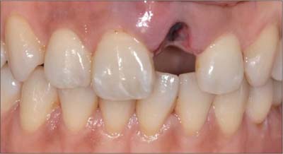
Figure 1 Intraoral photograph of avulsed maxillary left central incisor.

Figure 2 Eight weeks after trauma. (a) Intraoral photograph (labial view); (b) Periapical view.

Figure 3 One year after trauma. (a) Intraoral photograph (labial view); (b) Intraoral photograph (palatal view); (c) Periapical view.

Figure 4 Palatal view of study model on CO bite. Minimal contact was visible.
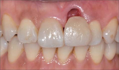
Figure 5 Temporary composite pontic was bonded after extraction of the maxillary left central incisor.
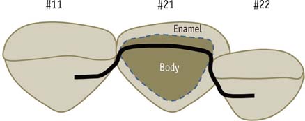
Figure 6 Diagram of the restoration, which shows how the FRC framework was placed.
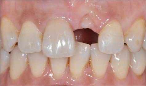
Figure 7 Two weeks after extraction of the maxillary left central incisor.
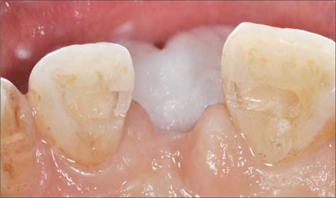
Figure 8 Palatal view after the abutment preparation (3 × 1 × 1 mm) was done. All margins were in the enamel, and the internal line angle was rounded.
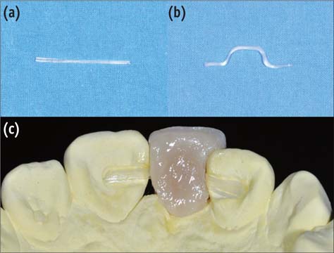
Figure 9 Prefabricated FRC framework and pontic. (a) FibreKor; (b) Light-cured FRC after forming the framework; (c) Pontic fabrication up to body layer.
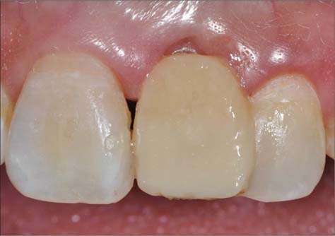
Figure 10 Prefabricated FRC fixed partial prosthesis was bonded to the abutment.
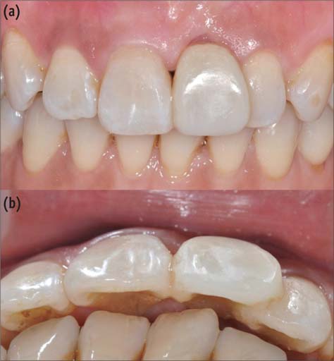
Figure 11 Postoperative view. (a) Labial view; (b) Occlusal view.

Figure 12 One-year recall. (a) Labial view; (b) Palatal view; (c) Periapical view.
Reference
-
1. Strassler HE, Serio CL. Esthetic considerations when splinting with fiber-reinforced composites. Dent Clin North Am. 2007; 51:507–524.
Article2. Eminkahyagil N, Erkut S. An innovative approach to chairside provisional replacement of an extracted anterior tooth: use of fiber-reinforced ribbon-composites and a natural tooth. J Prosthodont. 2006; 15:316–320.
Article3. Byun CW, Park SH, Park SJ, Choi KK. The effect of reinforcing methods on fracture strength of composite inlay bridge. J Korean Acad Conserv Dent. 2007; 32:111–120.
Article4. Chafaie A, Portier R. Anterior fiber-reinforced composite resin bridge: a case report. Pediatr Dent. 2004; 26:530–534.5. Kermanshah H, Motevasselian F. Immediate tooth replacement using fiber-reinforced composite and natural tooth pontic. Oper Dent. 2010; 35:238–245.
Article6. Zarow M, Paisley CS, Krupinski J, Brunton PA. Fiber-reinforced composite fixed dental prostheses: two clinical reports. Quintessence Int. 2010; 41:471–477.7. Kim KY, Kim SY, Kim DS, Choi KK. Use of temporary filling material for index fabrication in Class IV resin composite restoration. Restor Dent Endod. 2013; 38:85–89.
Article8. Song HY, Yi YJ, Cho LR, Park DY. Effects of two preparation designs and pontic distance on bending and fracture strength of fiber-reinforced composite inlay fixed partial dentures. J Prosthet Dent. 2003; 90:347–353.
Article9. Shinya A, Yokoyama D, Lassila LV, Shinya A, Vallittu PK. Three-dimensional finite element analysis of metal and FRC adhesive fixed dental prostheses. J Adhes Dent. 2008; 10:365–371.10. Salama H, Salama MA, Garber D, Adar P. The interproximal height of bone: a guidepost to predictable aesthetic strategies and soft tissue contours in anterior tooth replacement. Pract Periodontics Aesthet Dent. 1998; 10:1131–1141.11. Freilich MA, Meiers JC, Duncan JP, Eckrote KA, Goldberg AJ. Clinical evaluation of fiber-reinforced fixed bridges. J Am Dent Assoc. 2002; 133:1524–1534.
Article12. Jain P, Cobb D. Evaluation of a glass-fiber-reinforced, bonded, inlay-supported fixed partial denture-4-year results. Compend Contin Educ Dent. 2002; 23:779–783.13. Vallittu PK. Survival rates of resin-bonded, glass fiber-reinforced composite fixed partial dentures with a mean follow-up of 42 months: a pilot study. J Prosthet Dent. 2004; 91:241–246.
Article14. Wolff D, Schach C, Kraus T, Ding P, Pritsch M, Mente J, Joerss D, Staehle HJ. Fiber-reinforced composite fixed dental prostheses: a retrospective clinical examination. J Adhes Dent. 2011; 13:187–194.
- Full Text Links
-
- Actions
-
Cited
- CITED
-
- Close
- Share
-
- Similar articles
-
- Currently there are so many fiber reinforced composite posts in the market. Some products are factory silanated but some products are not. Should I use silane for surface treatment of fiber reinforced composite posts?
- Fiber-reinforced composite resin bridges: an alternative method to treat root-fractured teeth
- The influence of fitness and type of luting agents on bonding strength of fiber-reinforced composite resin posts
- Effect of fiber direction on the polymerization shrinkage of fiber-reinforced composites
- Comparison between fiber-reinforced polymers and stainless steel orthodontic retainers
