Misdiagnosis of florid cemento-osseous dysplasia leading to unnecessary root canal treatment: a case report
- Affiliations
-
- 1Department of Oral and Maxillofacial Surgery, Yonsei University College of Dentistry, Seoul, Korea.
- 2Department of Conservative Dentistry, Yonsei University College of Dentistry, Seoul, Korea. shujungshin@yahoo.com
Abstract
- This case report demonstrates an unnecessary endodontic treatment of teeth with florid cemento-osseous dysplasia (FCOD) due to a misdiagnosis as periapical pathosis and emphasizes the importance of correct diagnosis to avoid unnecessary treatment. A 30-year-old woman was referred to our institution for apicoectomies of the mandibular left canine and both the lateral incisors. The periapical lesions associated with these teeth had failed to resolve after root canal treatment over a 3-year period. Radiographic examinations revealed multiple lesions on the right canine, the second premolar, and both first molars as well as the anterior region of the mandible. Based on clinical, radiographic and histological evaluations, the patient condition was diagnosed as FCOD. The patient has been monitored for 2 years. To avoid unnecessary invasive treatment, accurate diagnosis is essential before treatment is carried out in managing FCOD.
Keyword
MeSH Terms
Figure
-

Figure 1 A series of panoramic radiographs obtained during a previous dental consultation. (a) A panoramic radiograph obtained in February, 2005 (4 years before the initial visit to our institution) when the patient's dentist initiated orthodontic treatment. Slight periapical radiolucency on #32, 33, and 42 was detected; (b) A panoramic radiograph obtained in October, 2005 when the root canal therapies were performed on #32, 33, and 42; (c) A panoramic radiograph obtained in 2007; (d) A panoramic radiograph obtained in 2008. Compared with the radiographs in 2005, the size of the lesion in the mandibular incisors increased, and a mixed radiopaque and radiolucent area was clearly distinguishable in the apical areas of the right first molar, right second molar, and left first molar.
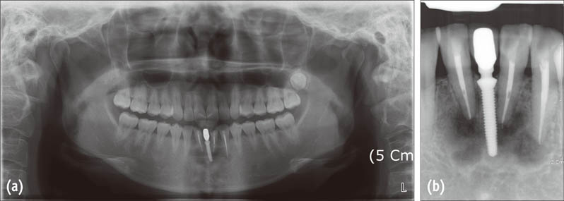
Figure 2 Radiographic examinations performed at the initial visit (in April, 2009) of the patient. (a) A panoramic radiograph showing multiple mixed radiopaque and radiolucent areas at the apices of the mandibular teeth; (b) A periapical radiograph of the lower anterior teeth. Two incisors and left canine were endodontically treated and an implant was included in the apical lesion.
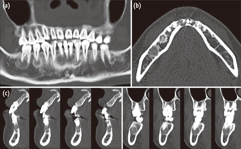
Figure 3 Computed tomography (CT) images at the initial visit. (a) A panoramic reconstruction of the CT images at the level of the apices. Multiple lesions bony lesions were detected; (b) Axial images of #42, 32, and 46 at the apical level. Mixed radiopaque and radiolucent lesions as well as cortical bone perforation were observed; (c) Vertical images showing thinning and perforation of the cortical plate without any expansion.
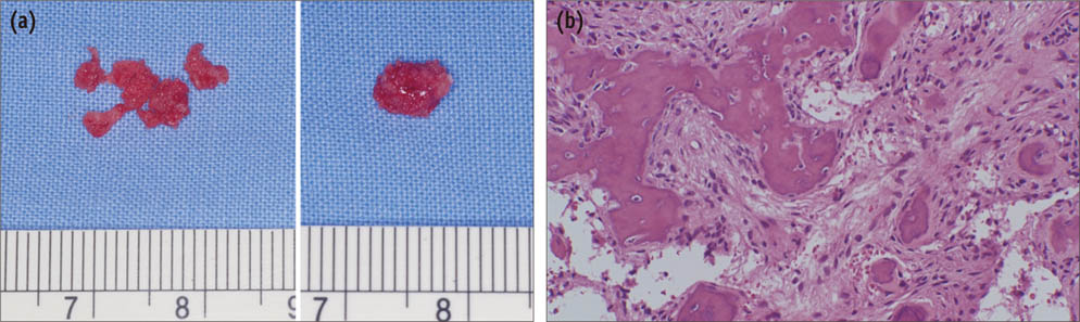
Figure 4 Results of examination of the excisional biopsy specimens. (a) Gross appearance of the specimens. The largest specimen was 7.0 × 5.0 × 5.0 mm; (b) A histologic examination showed bony trabeculae mixed with cellularity in the stroma (hematoxylin-eosin stain, ×50).
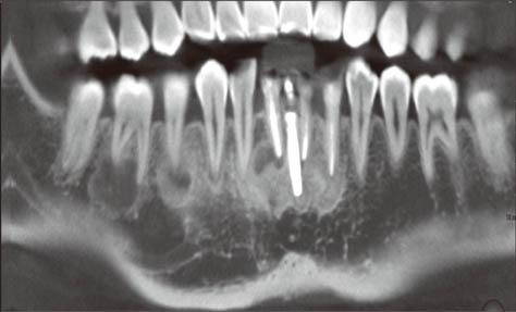
Figure 5 Radiographic examinations performed in 2011. A panoramic reconstructed image of cone-beam computed tomography (CBCT) obtained in September, 2011 (21 months after the biopsy).
Reference
-
1. Waldron CA. Bone pathology. In : Neville BW, Damm DD, Allen CM, Bouquot JE, editors. Oral & Maxillofacial Pathology. 2nd ed. Philadelphia: Saunders;2002. p. 533–587.2. Beylouni I, Farge P, Mazoyer JF, Coudert JL. Florid cemento-osseous dysplasia: report of a case documented with computed tomography and 3D imaging. Oral Surg Oral Med Oral Pathol Oral Radiol Endod. 1998; 85:707–711.3. Kramer IR, Pindborg JJ, Shear M. The WHO histological typing of odontogenic tumours. A commentary on the second edition. Cancer. 1992; 70:2988–2994.
Article4. MacDonald-Jankowski DS. Florid cemento-osseous dysplasia: a systematic review. Dentomaxillofac Radiol. 2003; 32:141–149.
Article5. Summerlin DJ, Tomich CE. Focal cemento-osseous dysplasia: a clinicopathologic study of 221 cases. Oral Surg Oral Med Oral Pathol. 1994; 78:611–620.
Article6. Su L, Weathers DR, Waldron CA. Distinguishing features of focal cemento-osseous dysplasia and cemento-ossifying fibromas. II. A clinical and radiologic spectrum of 316 cases. Oral Surg Oral Med Oral Pathol Oral Radiol Endod. 1997; 84:540–549.
Article7. Su L, Weathers DR, Waldron CA. Distinguishing features of focal cemento-osseous dysplasias and cemento-ossifying fibromas: I. A pathologic spectrum of 316 cases. Oral Surg Oral Med Oral Pathol Oral Radiol Endod. 1997; 84:301–309.
Article8. Mupparapu M, Singer SR, Milles M, Rinaggio J. Simultaneous presentation of focal cemento-osseous dysplasia and simple bone cyst of the mandible masquerading as a multilocular radiolucency. Dentomaxillofac Radiol. 2005; 34:39–43.
Article9. Rodrigues CD, Estrela C. Periapical cemento-osseous dysplasia in maxillary teeth suggesting apical periodontitis: case report. Gen Dent. 2009; 57:e21–e24.10. Galgano C, Samson J, Küffer R, Lombardi T. Focal cemento-osseous dysplasia involving a mandibular lateral incisor. Int Endod J. 2003; 36:907–911.
Article11. Resnick CM, Novelline RA. Cemento-osseous dysplasia, a radiological mimic of periapical dental abscess. Emerg Radiol. 2008; 15:367–374.
Article12. Kawai T, Hiranuma H, Kishino M, Jikko A, Sakuda M. Cemento-osseous dysplasia of the jaws in 54 Japanese patients: a radiographic study. Oral Surg Oral Med Oral Pathol Oral Radiol Endod. 1999; 87:107–114.13. Komabayashi T, Zhu Q. Cemento-osseous dysplasia in an elderly Asian male: a case report. J Oral Sci. 2011; 53:117–120.
Article14. Miyake M, Nagahata S. Florid cemento-osseous dysplasia. Report of a case. Int J Oral Maxillofac Surg. 1999; 28:56–57.
Article15. Ong ST, Siar CH. Florid cemento-osseous dysplasia in a young Chinese man. Case report. Aust Dent J. 1997; 42:404–408.
Article16. Cavalcante AS, Sgarbi FC, Agapito Lda C, Roveroni LH, Brandao AA, Cabral LA. Florid cemento-osseous dysplasia: a report of three cases. Gen Dent. 2008; 56:186–190.17. Singer SR, Mupparapu M, Rinaggio J. Florid cemento-osseous dysplasia and chronic diffuse osteomyelitis: report of a simultaneous presentation and review of the literature. J Am Dent Assoc. 2005; 136:927–931.18. Islam MN, Cohen DM, Kanter KG, Stewart CM, Katz J, Bhattacharyya I. Florid cemento-osseous dysplasia mimicking multiple periapical pathology-an endodontic dilemma. Gen Dent. 2008; 56:559–562.19. Makek MS. So called 'fibro-osseous lesions' of tumorous origin. Biology confronts terminology. J Craniomaxillofac Surg. 1987; 15:154–167.20. Ariji Y, Ariji E, Higuchi Y, Kubo S, Nakayama E, Kanda S. Florid cemento-osseous dysplasia. Radiographic study with special emphasis on computed tomography. Oral Surg Oral Med Oral Pathol. 1994; 78:391–396.21. Alsufyani NA, Lam EW. Cemento-osseous dysplasia of the jaw bones: key radiographic features. Dentomaxillofac Radiol. 2011; 40:141–146.
Article22. Bencharit S, Schardt-Sacco D, Zuniga JR, Minsley GE. Surgical and prosthodontic rehabilitation for a patient with aggressive florid cemento-osseous dysplasia: a clinical report. J Prosthet Dent. 2003; 90:220–224.
Article
- Full Text Links
-
- Actions
-
Cited
- CITED
-
- Close
- Share
-
- Similar articles
-
- 3 Types of Cemento-Osseous Dysplasia: Case Reports
- Florid cemento-osseous dysplasia: a report of two cases
- Recurrent symptomatic cemento-osseous dysplasia: A case report
- Clinical, radiographic, and histological findings of florid cemento-osseous dysplasia: a case report
- Bisphosphonate-Related Osteonecrosis in a Patient with Florid Cemento-Osseous Dysplasia
