Brain and Skin Metastasis From Urothelial Carcinoma of the Bladder
- Affiliations
-
- 1Department of Urology, Hanyang University College of Medicine, Seoul, Korea. qpqp@hanyang.ac.kr
- 2Department of Pathology, Hanyang University College of Medicine, Seoul, Korea.
Abstract
- Brain and skin metastasis from urothelial carcinoma of the bladder is rare. There have been few case reports of the clinical course of patients with metastatic urothelial carcinoma of the brain and skin. In the present case, a 60-year-old man had undergone radical cystectomy with an ileal conduit owing to urothelial carcinoma (T1N0M0). The patient developed dizziness 9 years later and a solitary brain tumor was discovered in his left cerebellar hemisphere. The tumor was totally resected and the mass was verified to be metastatic urothelial carcinoma. One year after the metastasectomy of the brain lesion, multiple erythematous nodular lesions developed on his abdominal skin. The skin lesions were excised and verified to be metastatic urothelial carcinoma. This report describes this case of urothelial carcinoma of the bladder that metastasized to the brain and abdominal skin.
Keyword
MeSH Terms
Figure
-
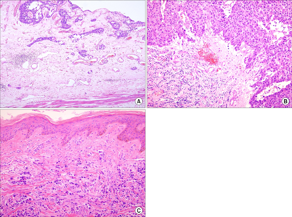
FIG. 1 (A) A representative section of the urinary bladder shows high-grade urothelial carcinoma. The tumor cells are arranged in small nests or cords and discontinuously invade into deep portions of the lamina propria (H&E, ×100). (B) The brain mass of the cerebellum shows metastatic carcinoma with tumor cells in a broad cord or sheet arrangement, identical to a urinary bladder tumor (H&E, ×200). (C) The excisional biopsied skin the abdomen shows individually scattered and nested pleomorphic tumor cells in the dermis, which is consistent with metastatic urothelial carcinoma (H&E, ×200).
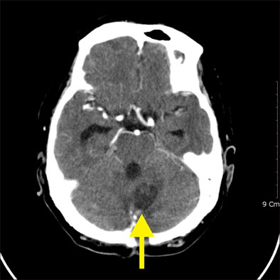
FIG. 2 Computed tomography shows a 3×4 cm sized mass (arrow) in the left cerebellar hemisphere.
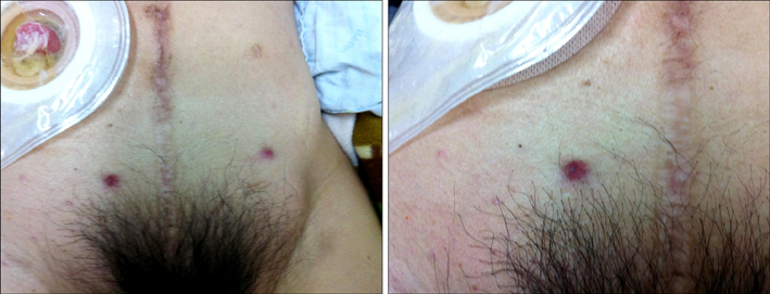
FIG. 3 Clinical pictures show multiple 1 cm sized erythematic nodular abdominal skin lesions.
Reference
-
1. Wakisaka S, Miyahara S, Nonaka A, Asami N, Kinoshita K, Kohriyama K. Brain metastasis from transitional cell carcinoma of the bladder: case report. Neurol Med Chir (Tokyo). 1990. 30:188–190.2. Swick BL, Gordon JR. Superficially invasive transitional cell carcinoma of the bladder associated with distant cutaneous metastases. J Cutan Pathol. 2010. 37:1245–1250.3. Atmaca AF, Akbulut Z, Demirci A, Belenli O, Alici S, Balbay DM. Multiple subcutaneous nodular metastases from transitional cell carcinoma of the bladder. Pathol Oncol Res. 2007. 13:70–72.4. Shinagare AB, Ramaiya NH, Jagannathan JP, Fennessy FM, Taplin ME, Van den Abbeele AD. Metastatic pattern of bladder cancer: correlation with the characteristics of the primary tumor. AJR Am J Roentgenol. 2011. 196:117–122.5. Babaian RJ, Johnson DE, Llamas L, Ayala AG. Metastases from transitional cell carcinoma of urinary bladder. Urology. 1980. 16:142–144.6. Goldman SM, Fajardo AA, Naraval RC, Madewell JE. Metastatic transitional cell carcinoma from the bladder: radiographic manifestions. AJR Am J Roentgenol. 1979. 132:419–425.7. Oh SH, Lee WJ, Rhee DY, Chang SE, Lee MW, Choi JH, et al. A case of cutaneous metastasis from urothelial carcinoma of the urinary bladder. Korean J Dermatol. 2007. 45:401–403.8. Ku JH, Yeo WG, Park MY, Lee ES, Kim HH. Metastasis of transitional cell carcinoma to the lower abdominal wall 20 years after cystectomy. Yonsei Med J. 2005. 46:181–183.9. Mueller TJ, Wu H, Greenberg RE, Hudes G, Topham N, Lessin SR, et al. Cutaneous metastases from genitourinary malignancies. Urology. 2004. 63:1021–1026.10. Spector JI, Zimbler H, DeLeo M, Ross JS. Skin metastases from transitional cell bladder cancer. Urology. 1987. 29:215–217.
- Full Text Links
-
- Actions
-
Cited
- CITED
-
- Close
- Share
-
- Similar articles
-
- A Case of Cutaneous Metastasis from Urothelial Carcinoma of the Urinary Bladder
- A Case of Cutaneous Metastasis from Carcinoma of the Urinary Bladder
- Skeletal Muscle Metastases from Urothelial Cell Carcinoma
- A Case of Cutaneous Metastasis from Carcinoma of the Urinary Bladder
- Seminal Vesicle Involvement by Urothelial Carcinoma in Situ of the Bladder with Mucosal Spread Pattern: A Case Report
