Pediatr Gastroenterol Hepatol Nutr.
2014 Jun;17(2):98-103.
Acalculous Diffuse Gallbladder Wall Thickening in Children
- Affiliations
-
- 1Postgraduate School of Medicine, Pusan National University, Yangsan, Korea.
- 2Department of Pediatrics, Pusan National University School of Medicine, Yangsan, Korea. jhongpark@pusan.ac.kr
- 3Department of Radiology, Pusan National University School of Medicine, Yangsan, Korea.
Abstract
- PURPOSE
Gallbladder (GB) wall thickening can be found in various conditions unrelated to intrinsic GB disease. We investigated the predisposing etiologies and the outcome of acalculous GB wall thickening in children.
METHODS
We retrospectively analyzed 67 children with acalculous GB wall thickening who had visited our institute from June 2010 to June 2013. GB wall thickening was defined as a GB wall diameter >3.5 mm on abdominal ultrasound examination or computed tomography. Underlying diseases associated with GB wall thickening, treatment, and outcomes were studied.
RESULTS
There were 36 boys and 31 girls (mean age, 8.5+/-4.8 years [range, 7 months-16 years]). Systemic infection in 24 patients (35.8%), acute hepatitis in 18 (26.9%), systemic disease in 11 (16.4%), hemophagocytic lymphohistiocytosis in 4 (6.0%), acute pancreatitis in 3 (4.5%), and specific liver disease in 3 (4.5%) predisposed patients to GB wall thickening. Systemic infections were caused by bacteria in 10 patients (41.7%), viruses in 5 patients (20.8%), and fungi in 2 patients (8.3%). Systemic diseases observed were systemic lupus erythematosus in 2, drug-induced hypersensitivity in 2, congestive heart failure in 2, renal disorder in 2. Sixty-one patients (91.0%) received symptomatic treatments or treatment for underlying diseases. Five patients (7.5%) died from underlying diseases. Cholecystectomy was performed in 3 patients during treatment of the underlying disease.
CONCLUSION
A wide range of extracholecystic conditions cause diffuse GB wall thickening that resolves spontaneously or with treatment of underlying diseases. Surgical treatments should be avoided if there are no definite clinical manifestations of cholecystitis.
MeSH Terms
Figure
-
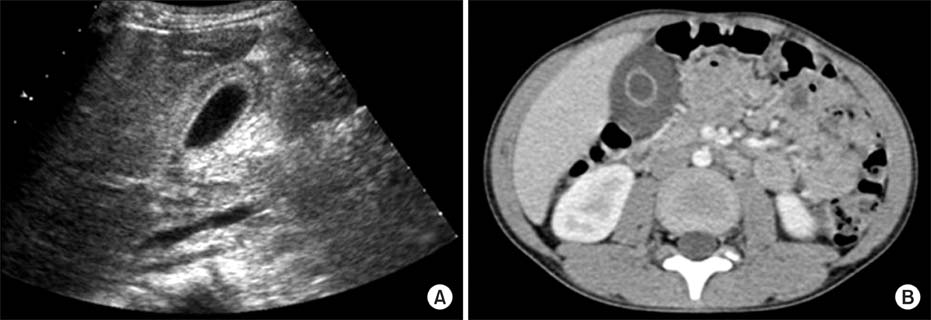
Fig. 1 An abdominal ultrasound sonography (A) and a computed tomography scan (B) showing a diffuse thickened gallbladder wall >3.5 mm in diameter.
Reference
-
1. van Breda Vriesman AC, Engelbrecht MR, Smithuis RH, Puylaert JB. Diffuse gallbladder wall thickening: differential diagnosis. AJR Am J Roentgenol. 2007; 188:495–501.
Article2. Tsakayannis DE, Kozakewich HP, Lillehei CW. Acalculous cholecystitis in children. J Pediatr Surg. 1996; 31:127–130. discussion 30-1.
Article3. Imamoğlu M, Sarihan H, Sari A, Ahmetoğlu A. Acute acalculous cholecystitis in children: Diagnosis and treatment. J Pediatr Surg. 2002; 37:36–39.
Article4. Glenn F. Acute acalculous cholecystitis. Ann Surg. 1979; 189:458–465.5. Sievert W, Vakil NB. Emergencies of the biliary tract. Gastroenterol Clin North Am. 1988; 17:245–264.
Article6. Mirvis SE, Vainright JR, Nelson AW, Johnston GS, Shorr R, Rodriguez A, et al. The diagnosis of acute acalculous cholecystitis: a comparison of sonography, scintigraphy, and CT. AJR Am J Roentgenol. 1986; 147:1171–1175.
Article7. Shetty PB, Broome DR. Sonographic analysis of gallbladder findings in Salmonella enteric fever. J Ultrasound Med. 1998; 17:231–237.
Article8. Lee AW, Proudfoot WH, Griffen WO Jr. Acalculous cholecystitis. Surg Gynecol Obstet. 1984; 159:33–35.9. Weeder RS, Bashant GH, Muir RW. Acute noncalculous cholecystitis associated with severe injury. Am J Surg. 1970; 119:729–732.
Article10. Munster AM, Goodwin MN, Pruitt BA Jr. Acalculous cholecystitis in burned patients. Am J Surg. 1971; 122:591–593.
Article11. Fabian TC, Hickerson WL, Mangiante EC. Posttraumatic and postoperative acute cholecystitis. Am Surg. 1986; 52:188–192.12. Lee SC, Tchah H, Na SY, Kim HS, Park HJ, Shin MK. Clinical and histopathologic findings on the abnormal liver function complicated with Kawasaki disease. Korean J Pediatr Gastroenterol Nutr. 2000; 3:47–55.
Article13. Patriquin HB, DiPietro M, Barber FE, Teele RL. Sonography of thickened gallbladder wall: causes in children. AJR Am J Roentgenol. 1983; 141:57–60.
Article14. Yamada K, Yamada H. Gallbladder wall thickening in mononucleosis syndromes. J Clin Ultrasound. 2001; 29:322–325.
Article15. Rumack CM, Wilson SR, Charboneau JW. Diagnostic ultrasound. 2nd ed. St. Louis: Mosby;1998.16. Kaftori JK, Pery M, Green J, Gaitini D. Thickness of the gallbladder wall in patients with hypoalbuminemia: a sonographic study of patients on peritoneal dialysis. AJR Am J Roentgenol. 1987; 148:1117–1118.
Article17. Maudgal DP, Wansbrough-Jones MH, Joseph AE. Gallbladder abnormalities in acute infectious hepatitis. A prospective study. Dig Dis Sci. 1984; 29:257–260.18. Giorgio A, Francica G, Amoroso P, Fico P, de Stefano G, Pierri P, et al. Morphologic and motility changes of the gallbladder in response to acute liver injury. A prospective real-time sonographic study in 255 patients with acute viral hepatitis. J Ultrasound Med. 1989; 8:499–506.
Article19. Suk KT, Kim CH, Baik SK, Kim MY, Park DH, Kim KH, et al. Gallbladder wall thickening in patients with acute hepatitis. J Clin Ultrasound. 2009; 37:144–148.
Article20. Kim MY, Baik SK, Choi YJ, Park DH, Kim HS, Lee DK, et al. Endoscopic sonographic evaluation of the thickened gallbladder wall in patients with acute hepatitis. J Clin Ultrasound. 2003; 31:245–249.
Article21. Schmidt MH, Sung L, Shuckett BM. Hemophagocytic lymphohistiocytosis in children: abdominal US findings within 1 week of presentation. Radiology. 2004; 230:685–689.
Article
- Full Text Links
-
- Actions
-
Cited
- CITED
-
- Close
- Share
-
- Similar articles
-
- Adequate Management of Gallbladder Wall Thickening
- A Case of Acute Acalculous Cholecystitis Superimposed on the Nephrotic Syndrome
- Age, Predisposing Diseases, and Ultrasonographic Findings in Determining Clinical Outcome of Acute Acalculous Inflammatory Gallbladder Diseases in Children
- Gallbladder Wall Thickening in Patients with Scrub Typhus: US Findings
- Gallbladder Wall Thickening and Periportal Tracking on CT in Patients with Acute Pyelonephritis
