Korean J Urol.
2012 Jun;53(6):386-390.
Clinical Outcomes of Bosniak Category IIF Complex Renal Cysts in Korean Patients
- Affiliations
-
- 1Department of Urology, Sahmyook Medical Center, Seoul, Korea.
- 2Urological Science Institute, Yonsei University College of Medicine, Seoul, Korea. uroham@yuhs.ac
Abstract
- PURPOSE
To assess the clinical reliability of the Bosniak IIF category and to determine the proper radiologic follow-up duration and intervals for category IIF complex renal cysts.
MATERIALS AND METHODS
We studied 201 patients with category IIF renal cysts from January 1996 to January 2011. Renal cyst progression to category III was defined as an increase in complexity of the cyst in follow-up radiologic studies. We monitored radiologic changes and progression of renal cysts during the follow-up period and analyzed the pathologic results of those patients who were treated surgically.
RESULTS
At a mean follow-up of 20 months, only 14 cases (7%) showed evidence of progression to stage III, with a mean time to progression of 11 months (range, 3 to 65 months). There were no significant differences in age, gender, cyst size, or change in cyst size between the progressive and non-progressive groups. Of 12 cases treated surgically, 10 cases (83.3%) showed renal cell carcinoma with pT1 stage, and there was no recurrence during postoperative follow-up of 23 months. Of the 187 patients without radiologic progression, 23 cases were treated surgically, and all of them showed benign cysts.
CONCLUSIONS
The IIF category showed significant clinical reliability by a low rate of radiologic progression and a high rate of malignancy in the radiologic progressive group but a low rate of malignancy in the non-progressive group. Although it is hard to decide on a proper follow-up duration because of the variable time to progression, too frequent follow-up study seems to be unnecessary considering that most malignant cases were of a low stage.
Keyword
Figure
-
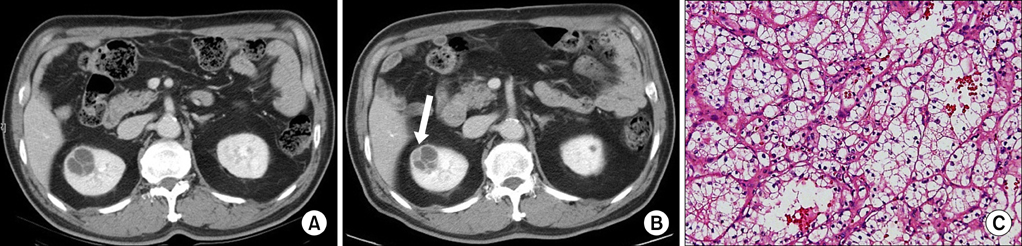
FIG. 1 A 69-year-old man with a category IIF cystic lesion in the right kidney. (A) Initial contrast-enhanced transverse computed tomography scan depicts a 2.8-cm cystic mass that contains multiple minimally smooth thickening of septa. (B) On follow-up 9 months later, the cystic renal mass contains grossly thickened and irregular septa in which there is measurable enhancement (white arrow). On the basis of these findings, the lesion was reclassified as a category III cyst. (C) The patient underwent radical nephrectomy and the lesion was found to be a pT1 stage conventional renal cell carcinoma with focal hemorrhage (H&E, ×400).
Reference
-
1. Kissane JM. The morphology of renal cystic disease. Perspect Nephrol Hypertens. 1976. 4:31–63.2. Kim JC. Usefulness of the Bosniak classification in cystic renal mass on CT. J Korean Radiol Soc. 1999. 40:555–562.3. Park HS, Jeong KS, Cheon J, Yoon DK, Jeong KB. The clinical significance of Bosniak classification in cystic renal masses: usefulness of preoperative computerized tomography in cystic renal masses. Korean J Urol. 1994. 35:498–503.4. Bosniak MA. The current radiological approach to renal cysts. Radiology. 1986. 158:1–10.5. Siegel CL, McFarland EG, Brink JA, Fisher AJ, Humphrey P, Heiken JP. CT of cystic renal masses: analysis of diagnostic performance and interobserver variation. AJR Am J Roentgenol. 1997. 169:813–818.6. Bosniak MA. Problems in the radiologic diagnosis of renal parenchymal tumors. Urol Clin North Am. 1993. 20:217–230.7. Bosniak MA. Diagnosis and management of patients with complicated cystic lesions of the kidney. AJR Am J Roentgenol. 1997. 169:819–821.8. O'Malley RL, Godoy G, Hecht EM, Stifelman MD, Taneja SS. Bosniak category IIF designation and surgery for complex renal cysts. J Urol. 2009. 182:1091–1095.9. Israel GM, Bosniak MA. Follow-up CT of moderately complex cystic lesions of the kidney (Bosniak category IIF). AJR Am J Roentgenol. 2003. 181:627–633.10. Graumann O, Osther SS, Osther PJ. Characterization of complex renal cysts: a critical evaluation of the Bosniak classification. Scand J Urol Nephrol. 2011. 45:84–90.11. Smith AD, Remer EM, Cox KL, Lieber ML, Allen BC, Shah SN, et al. Bosniak category IIF and III cystic renal lesions: outcomes and associations. Radiology. 2012. 262:152–160.12. Curry NS, Cochran ST, Bissada NK. Cystic renal masses: accurate Bosniak classification requires adequate renal CT. AJR Am J Roentgenol. 2000. 175:339–342.13. Bielsa O, Lloreta J, Gelabert-Mas A. Cystic renal cell carcinoma: pathological features, survival and implications for treatment. Br J Urol. 1998. 82:16–20.14. Smaldone MC, Uzzo RG. Active surveillance: a potential strategy for select patients with small renal masses. Future Oncol. 2011. 7:1133–1147.15. Eble JN, Bonsib SM. Extensively cystic renal neoplasms: cystic nephroma, cystic partially differentiated nephroblastoma, multilocular cystic renal cell carcinoma, and cystic hamartoma of renal pelvis. Semin Diagn Pathol. 1998. 15:2–20.16. Murad T, Komaiko W, Oyasu R, Bauer K. Multilocular cystic renal cell carcinoma. Am J Clin Pathol. 1991. 95:633–637.
- Full Text Links
-
- Actions
-
Cited
- CITED
-
- Close
- Share
-
- Similar articles
-
- Differential Diagnosis of Complex Renal Cysts Based on Lesion Size along with the Bosniak Renal Cyst Classification
- Usefulness of the Bosniak Classification in Cystic Renal Mass on CT
- The Clinical Significance of Bosniak Classification in Cystic Renal Masses : Usefulness of Preoperative Computerized Tomography in Cystic Renal Masses
- The role of Bosniak classification in malignant tumor diagnosis: A single institution experience
- Category Migration of Renal Cystic Masses with Use of Gadolinium-Enhanced Magnetic Resonance Imaging
