Pediatr Allergy Respir Dis.
2011 Dec;21(4):350-355.
A Case of Mediastinal and Pulmonary Cryptococcosis in a 3-Year-Old Immunocompetent Girl
- Affiliations
-
- 1Department of Pediatrics, Institute of Allergy, Yonsei University College of Medicine, Seoul, Korea. mhsohn@yuhs.ac
- 2Department of Diagnostic Radiology, Yonsei University College of Medicine, Seoul, Korea.
- 3Department of Pathology, CHA University College of Medicine, Pocheon, Korea.
Abstract
- Cryptococcosis is an infrequently recognized infection in children, particularly those who are immunocompetent. The disease is mainly caused by Cryptococcus neoformans, a fungal pathogen that primarily affects the central nervous system (CNS) and lungs. Most reports of children with cryptococcosis are of the CNS or disseminated infections among immunocompromised patients. This report is a case of a 3-year-old immunocompetent girl who presented with intermittent fever and cough; a large mass was found in the right infrahilar area on chest X-ray. Chest computed tomography revealed large conglomerated mediastinal lymph nodes caused by C. neoformans, which was confirmed by the polymerase chain reaction as well as a histological evaluation. The patient improved after a prolonged period of antifungal therapy. This is the only known report of mediastinal and pulmonary cryptococcosis in an immunocompetent child.
Keyword
MeSH Terms
Figure
-

Fig. 1. Chest radiograph showing mediastinal widening and mass-like opacity (arrows) in the right lower lung field.
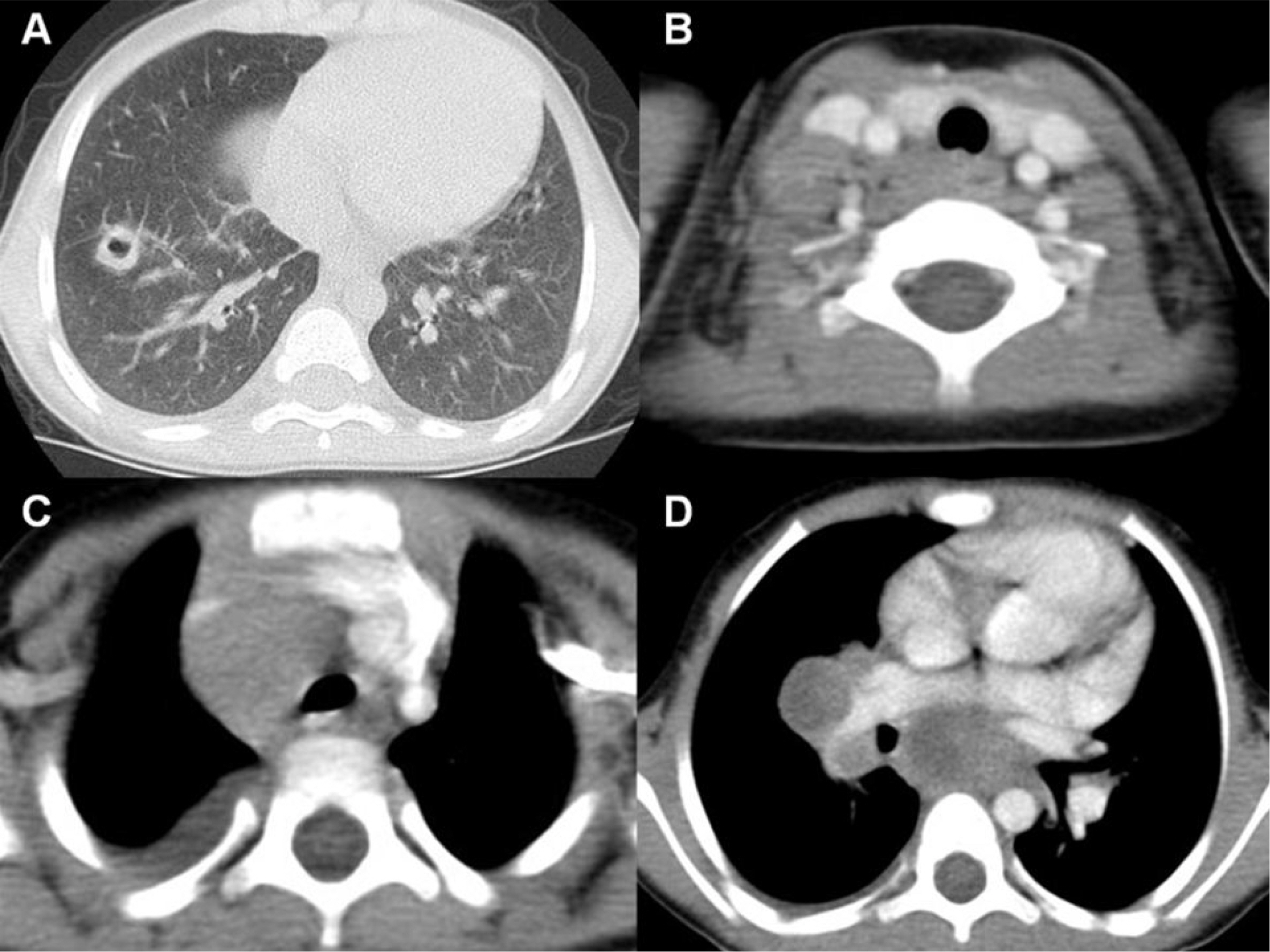
Fig. 2. Chest computed tomography scan demonstrating a cavitary lesion in the right lower lobe (A) with lymph node enlargement in the right lower neck (B), mediastinum (C), and hilum (D).
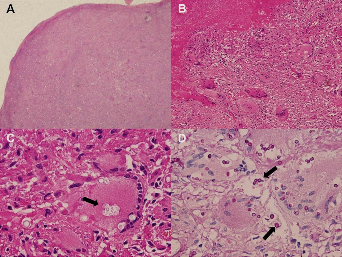
Fig. 3. Histopathology of the neck lymph node is consistent with Cryptococcus neoformans. Necrotizing granulomatous inflammation (A, H&E, 10; B, H&E, 100). Mucicarmine × × -positive yeasts of variable sizes (arrow) (C, H&E, 400). Yeasts with a thickened × capsule on periodic acid-Schiff stain (arrows) (D, 400). ×
Reference
-
References
1. Sweeney DA, Caserta MT, Korones DN, Casadevall A, Goldman DL. A ten-year-old boy with a pulmonary nodule secondary to Cryptococcus neoformans: case report and review of the literature. Pediatr Infect Dis J. 2003; 22:1089–93.
Article2. Chang WC, Tzao C, Hsu HH, Lee SC, Huang KL, Tung HJ, et al. Pulmonary cryptococcosis: comparison of clinical and radiographic characteristics in immunocompetent and immunocompromised patients. Chest. 2006; 129:333–40.3. Leggiadro RJ, Barrett FF, Hughes WT. Extrapulmonary cryptococcosis in immunocompromised infants and children. Pediatr Infect Dis J. 1992; 11:43–7.
Article4. Severo CB, Xavier MO, Gazzoni AF, Severo LC. Cryptococcosis in children. Paediatr Respir Rev. 2009; 10:166–71.
Article5. Jean SS, Fang CT, Shau WY, Chen YC, Chang SC, Hsueh PR, et al. Cryptococcaemia: clinical features and prognostic factors. QJM. 2002; 95:511–8.
Article6. Chaudhary MW, Sardana K, Kumar P, Dewan V, Anand VK. Disseminated infection with Cryptococcus neoformans var neoformans in an 8 years immunocompetent girl. Indian J Pediatr. 2005; 72:85.7. Nadrous HF, Antonios VS, Terrell CL, Ryu JH. Pulmonary cryptococcosis in nonimmunocom-promised patients. Chest. 2003; 124:2143–7.
Article8. Himmel JE, Stark P. Cryptococcal pneumonia in an immunocompetent host: radiographic findings. Semin Respir Infect. 2003; 18:129–31.9. Huang KY, Huang YC, Hung IJ, Lin TY. Cryptococcosis in nonhuman immunodeficiency virus-infected children. Pediatr Neurol. 2010; 42:267–70.
Article10. Yuanjie Z, Jianghan C, Julin G, Hai W. Cryptococcal meningitis in an immunocompetent 8-year-old girl. Mycoses. 2009; 52:377–8.
Article11. Vawda F, Maharajh J, Naidoo K. Massive cryptococcal lymphadenopathy in an immunocompetent pregnant patient. Br J Radiol. 2008; 81:e53–6.
Article12. Sinha P, Naik KG, Bhagwat GP. Mediastinal cryptococcoma. Thorax. 1978; 33:657–9.
Article13. Zou CC, Yu ZS, Tang LF, Liang L, Zhao ZY. Primary abdominal lymphonodular cryptococcosis in children: 2 case reports and a literature review. J Pediatr Surg. 2006; 41:e11–5.
Article14. Rozenbaum R, Gonçalves AJ. Clinical epidemiological study of 171 cases of cryptococcosis. Clin Infect Dis. 1994; 18:369–80.
Article15. Jarvis JN, Harrison TS. Pulmonary cryptococcosis. Semin Respir Crit Care Med. 2008; 29:141–50.
Article16. Lindell RM, Hartman TE, Nadrous HF, Ryu JH. Pulmonary cryptococcosis: CT findings in immunocompetent patients. Radiology. 2005; 236:326–31.
Article17. Sun LM, Chen TY, Chen WJ, Hsieh MJ, Liu JW, Huang CC, et al. Cryptococcus infection in a patient with nasopharyngeal carcinoma: imaging findings mimicking pulmonary metastases. Br J Radiol. 2002; 75:275–8.
Article18. Núñez M, Peacock JE Jr, Chin R Jr. Pulmonary cryptococcosis in the immunocompetent host. Therapy with oral fluconazole: a report of four cases and a review of the literature. Chest. 2000; 118:527–34.19. Dromer F, Mathoulin S, Dupont B, Brugiere O, Letenneur L. Comparison of the efficacy of amphotericin B and fluconazole in the treatment of cryptococcosis in human immunodeficiency virus-negative patients: retrospective analysis of 83 cases. French Cryptococcosis Study Group. Clin Infect Dis. 1996; 22(Suppl 2):S154–60.20. Perfect JR, Dismukes WE, Dromer F, Goldman DL, Graybill JR, Hamill RJ, et al. Clinical practice guidelines for the management of cryptococcal disease: 2010 update by the Infectious Diseases Society of America. Clin Infect Dis. 2010; 50:291–322.
Article
- Full Text Links
-
- Actions
-
Cited
- CITED
-
- Close
- Share
-
- Similar articles
-
- Massive Mediastinal Lymph Node Involvement of Cryptococcosis in Immunocompetent Host
- Cryptococcosis with Mediastinal Lymph Node and Lung Involvement in an Immunocompetent Adolescent: A Case Report
- HRCT Finding of Pulmonary Cryptococcosis in Immune Competent Patients
- Various Pulmonary Manifestations of the Cryptococcal Pneumoniae in the three Immunocompetent Patients
- Isolated pulmonary cryptococcosis in an immunocompetent boy
