Korean J Urol.
2010 Mar;51(3):212-215.
Life-Threatening Complication after Extracorporeal Shock Wave Lithotripsy for a Renal Stone: A Hepatic Subcapsular Hematoma
- Affiliations
-
- 1Department of Urology, Gachon University Gil Hospital, Incheon, Korea. kimcho99@gilhospital.com
Abstract
- Extracorporeal shock wave lithotripsy (ESWL) has revolutionized the management of urolithiasis since it was first introduced in 1980. ESWL is a well-established, safe and effective therapeutic alternative to surgical treatment for urolithiasis. Complications of ESWL do occur in a small number of patients, and when they do, they typically involve the kidney. We present a case of a young female patient who developed a huge hepatic subcapsular hematoma accompanied by hypovolemic shock after ESWL for a 9 mm stone in the right kidney. The hematoma measured 13x6 cm. Conservative care with no surgical intervention was chosen because there was no evidence of active bleeding on the computed tomography. After conservative therapy, the hematoma was gradually absorbed and the patient was discharged.
Keyword
Figure
-
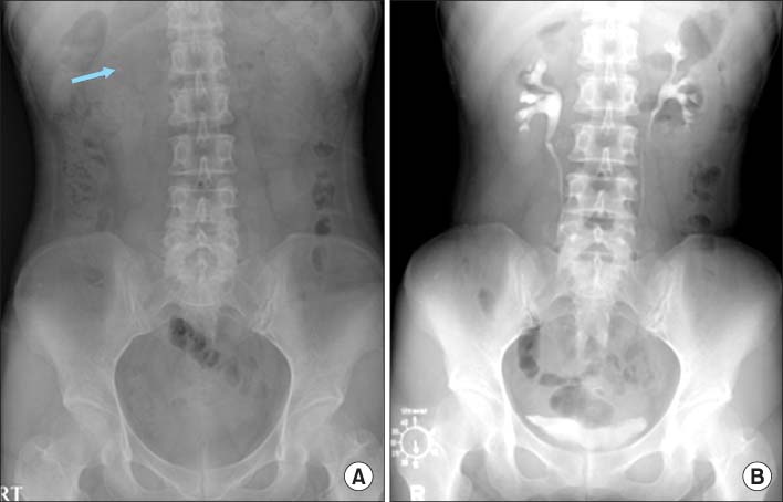
FIG. 1 (A) Before extracorporeal shock wave lithotripsy (ESWL), the kidney, ureter, and bladder radiography (KUB) revealed a 9 mm renal stone in the right kidney (arrow). (B) Before ESWL, the intravenous pyelography (IVP) revealed no hydronephrosis or excretion abnormality.

FIG. 2 (A) The axial and (B) vertical images of the computed tomography (CT) showing a large (13×6 cm) subcapsular hepatic hematoma.
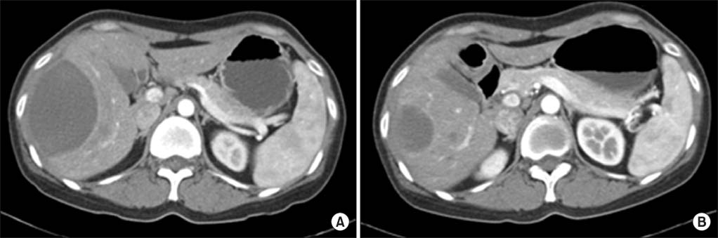
FIG. 3 (A) Two months after extracorporeal shock wave lithotripsy (ESWL), the CT scan revealed a decreased size of the subcapsular hepatic hematoma. (B) Five months after ESWL, the subcapsular hematoma had been markedly resorbed and presented in a resolving state.
Reference
-
1. Moon YT, Oh CH, Moon WC, Kim KD, Kim YS, Kim SC, et al. An experience with piezoelectric extracorporeal shock wave lithotripsy: 2000 cases. Korean J Nephrol. 1991. 10:166–174.2. Donahue LA, Linke CA, Rowe JM. Renal loss following extracorporeal shock wave lithotripsy. J Urol. 1989. 142:809–811.3. Kostakopoulos A, Stavropoulos NJ, Macrychoritis C, Deliveliotis C, Antonopoulos KP, Picramenos D. Subcapsular hematoma due to ESWL: risk factors. A study of 4,247 patients. Urol Int. 1995. 55:21–24.4. Sherman SC, Dogon A. Subcapsular renal hematoma after shock wave lithotripsy. J Emerg Med. 2006. 30:437–439.5. Beatrice J, Strebel RT, Pfammatter T, Röhweder JH, Sulser T. Life-threatening complication after right renal extracorporeal shock wave lithotripsy: large hepatic haematoma requiring embolisation of the right hepatic artery. Eur Urol. 2007. 52:909–911.6. Hirata N, Kushida Y, Ohguri T, Wakasugi S, Kojima T, Fujita R. Heptic subcapsular hematoma after extracorporeal shock wave lithotripsy (ESWL) for pancreatic stones. J Gastroenterol. 1999. 34:713–716.7. Gordetsky J, Hislop S, Orloff M, Butler M, Erturk E. Subcapsular hepatic hematoma with right hepatic vein thrombosis: a complication of shock wave lithotripsy. Can Urol Assoc J. 2008. 2:61–63.8. Klára T, Magdolna K. Fetal renal hemorrhage after extracorporeal shock wave lithotripsy. J Forensic. 2008. 53:1191–1193.9. Hirata N, Kushida Y, Ohguri T, Wakasugi S, Kojima T, Fujita R. Hepatic subcapsular hematoma after extracorpeal shock wave lithotripsy (ESWL) for pancreatic stones. J Gastroenterol. 1999. 34:713–716.10. Collado Serra A, Huguet Pérez J, Monreal García de Vicuña F, Rousaud Barón A, Izquierdo de la Torre F, Vicente Rodríguez J. Renal hematoma as a complication of extracorporeal shock wave lithotripsy. Scand J Urol Nephrol. 1999. 33:171–175.
- Full Text Links
-
- Actions
-
Cited
- CITED
-
- Close
- Share
-
- Similar articles
-
- Effects of Extracorporeal Shock Wave Lithortripsy Experimentally Induced Cholelithiasis and Organs in the Dog
- Clinical Experience of Extracorporeal Shock Wave Lithotripsy for Urinary Calculi
- Initial Clinical Experience of Extracorporeal Shock Wave Lithotripsy using the Northgate SD-3 Lithotriptor
- Extracorporeal shock wave lithotripsy of lower caliceal stone
- Use of Renal Scan(DTPA) for Clinical Follow-up of Renal Function after Extracorporeal Shock Wave Lithotripsy of Renal Stones
