Urachal Actinomycosis Mimicking a Urachal Tumor
- Affiliations
-
- 1Department of Urology, College of Medicine, Hanyang University, Seoul, Korea. moonuro@hanyang.ac.kr
- 2Department of Nuclear Medicine, College of Medicine, Hanyang University, Seoul, Korea.
- 3Department of Pathology, College of Medicine, Hanyang University, Seoul, Korea.
Abstract
- A 26-year-old man presented with lower abdominal discomfort and a palpable mass in the right lower quadrant. An abdominal computed tomography (CT) scan revealed an abdominal wall mass that extended from the dome of the bladder. Fluorine-18 fluorodeoxyglucose (FDG) positron-emission tomography/CT (PET/CT) showed hypermetabolic wall thickening around the bladder dome area that extended to the abdominal wall and hypermetabolic mesenteric infiltration. Differential diagnosis included a urachal tumor with invasion into adjacent organs and chronic inflammatory disease. Partial cystectomy with abdominal wall mass excision was performed, and the final pathologic report was consistent with urachal actinomycosis.
MeSH Terms
Figure
-
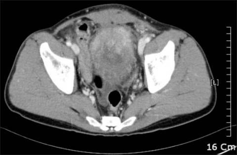
FIG. 1 Abdominal computed tomography (CT) showing wall thickening and a mass on the bladder dome. The mass extends to the abdominal wall from the bladder dome.
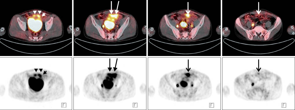
FIG. 2 Serial positron-emission tomography/computed tomography (PET/CT) images showing mild hypermetabolic wall thickening in the anterosuperior portion of the bladder (arrow heads) with hypermetabolic mesenteric and omental infiltration (arrows) extending along the urachus (thin arrow).
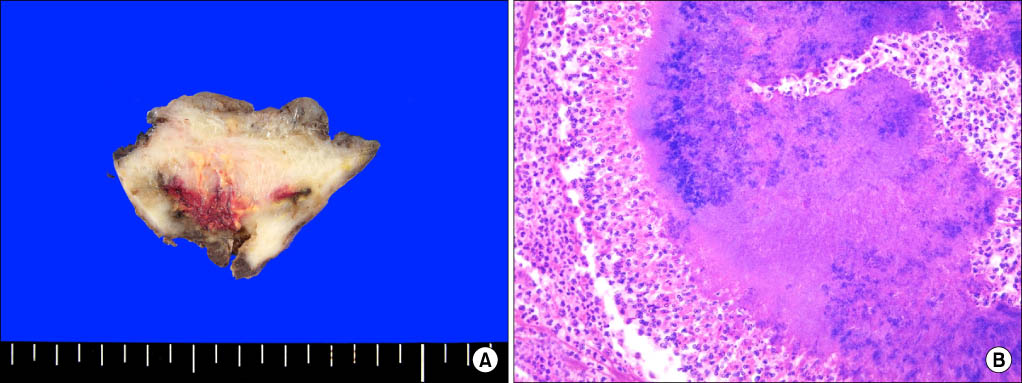
FIG. 3 (A) Partial cystectomy specimen revealing diffuse fibrotic thickening of the wall and relatively preserved luminal surface. There is an irregular necrotic lesion with yellowish foci in the deep resection margin. (B) Bacterial colony in the abscess background showing central entangled filamentous bacteria with a sunray appearance in the peripheral portion (H&E, ×400).
Reference
-
1. Choi CK, Rim HK, Kim JS, Rim JS. A case of urachal actinomycosis. Korean J Urol. 2000. 41:183–186.2. Lee SH, Kim KH, Kim JS, Kim MW, Lee HM, Lee KH, et al. A case of actinomycosis involving urachal remnant. Korean J Urol. 1999. 40:1714–1716.3. Lee HJ, Oh GS, Seo JB, Chung MK. Urachal Actinomycosis: report of a case. Korean J Urol. 1999. 40:933–936.4. Hsu CH, Lee CM, Chia CF, Lin YH. F-18 fluorodeoxyglucose positron emission tomography in an anorectal fistula with actinomycosis. Clin Nucl Med. 2004. 29:452–453.5. Yegüez JF, Martinez SA, Sands LR, Hellinger MD. Pelvic actinomycosis presenting as malignant large bowel obstruction: a case report and a review of the literature. Am Surg. 2000. 66:85–90.6. Cintron JR, Del Pino A, Duarte B, Wood D. Abdominal actinomycosis. Dis Colon Rectum. 1996. 39:105–108.7. Hsiao HL, Shen JT, Yeh HC, Wu WJ, Wang CJ, Huang CH. Intra- and extra-abdominal actinomycosis mimicking urachal tumor in an intrauterine device carrier: a case report. Kaohsiung J Med Sci. 2008. 24:35–40.8. Ho L, Seto J, Jadvar H. Actinomycosis mimicking anastomotic recurrent esophageal cancer on PET-CT. Clin Nucl Med. 2006. 31:646–647.9. Yeung VH, Wong QH, Chao NS, Leung MW, Kwok WK. Thoracic actinomycosis in an adolescent mimicking chest wall tumor or pulmonary tuberculosis. Pediatr Surg Int. 2008. 24:751–754.10. Al-Kadhi S, Venkiteswaran KP, Al-Ansari A, Shamsudini A, Al-Bozom I, Kiliyanni AS. Primary vesical actinomycosis: a case report and literature review. Int J Urol. 2007. 14:969–971.
