Obstet Gynecol Sci.
2015 Mar;58(2):144-149. 10.5468/ogs.2015.58.2.144.
Decreased bone mineral density is associated with coronary atherosclerosis in healthy postmenopausal women
- Affiliations
-
- 1Department of Obstetrics and Gynecology, Severance Hospital, Yonsei University College of Medicine, Seoul, Korea. dr222@yuhs.ac
- 2Institute of Women's Life Medical Science, Seoul, Korea.
- KMID: 2314038
- DOI: http://doi.org/10.5468/ogs.2015.58.2.144
Abstract
OBJECTIVE
This study aimed to assess the association between bone mineral density (BMD) and coronary atherosclerosis in healthy postmenopausal women.
METHODS
We performed a retrospective review of 252 postmenopausal women who had visited a health promotion center for a routine checkup. BMD of the lumbar spine (L1-L4) and femoral neck was evaluated using dual-energy X-ray absorptiometry, and coronary atherosclerosis was assessed using 64-row multidetector computed tomography. Participants were divided into normal BMD and osteopenia-osteoporosis groups, according to the T-scores of their lumbar spine or femoral neck.
RESULTS
Participants with osteopenia-osteoporosis had a significantly higher proportion of coronary atherosclerosis than did those with normal BMD at the lumbar spine (P=0.003) and femoral neck (P=0.004). Osteopenia-osteoporosis at the lumbar spine (odds ratio [OR], 2.86; 95% confidence interval [CI], 1.12 to 7.27) or femoral neck (OR, 3.35; 95% CI, 1.07 to 10.57) was associated with coronary atherosclerosis, after controlling for age and cardiovascular risk factors.
CONCLUSION
Decreased BMD is associated with coronary atherosclerosis in healthy postmenopausal women, independent of age and cardiovascular risk factors. Postmenopausal women with decreased BMD may have a higher risk of developing coronary atherosclerosis.
Keyword
MeSH Terms
Figure
-
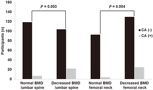
Fig. 1 Distribution of CA (-) and CA (+) patients in normal and osteopenia-osteoporosis groups. CA, coronary atherosclerosis; BMD, bone mineral density.
Reference
-
1. Von der Recke P, Hansen MA, Hassager C. The association between low bone mass at the menopause and cardiovascular mortality. Am J Med. 1999; 106:273–278.2. Witteman JC, Kok FJ, van Saase JL, Valkenburg HA. Aortic calcification as a predictor of cardiovascular mortality. Lancet. 1986; 2:1120–1122.3. Frye MA, Melton LJ 3rd, Bryant SC, Fitzpatrick LA, Wahner HW, Schwartz RS, et al. Osteoporosis and calcification of the aorta. Bone Miner. 1992; 19:185–194.4. Hak AE, Pols HA, van Hemert AM, Hofman A, Witteman JC. Progression of aortic calcification is associated with metacarpal bone loss during menopause: a population-based longitudinal study. Arterioscler Thromb Vasc Biol. 2000; 20:1926–1931.5. Kenchaiah S, Evans JC, Levy D, Wilson PW, Benjamin EJ, Larson MG, et al. Obesity and the risk of heart failure. N Engl J Med. 2002; 347:305–313.6. Browner WS, Seeley DG, Vogt TM, Cummings SR. Study of Osteoporotic Fractures Research Group. Nontrauma mortality in elderly women with low bone mineral density. Lancet. 1991; 338:355–358.7. Tanko LB, Bagger YZ, Christiansen C. Low bone mineral density in the hip as a marker of advanced atherosclerosis in elderly women. Calcif Tissue Int. 2003; 73:15–20.8. Seo SK, Cho S, Kim HY, Choi YS, Park KH, Cho DJ, et al. Bone mineral density, arterial stiffness, and coronary atherosclerosis in healthy postmenopausal women. Menopause. 2009; 16:937–943.9. Genant HK, Cooper C, Poor G, Reid I, Ehrlich G, Kanis J, et al. Interim report and recommendations of the World Health Organization Task-Force for Osteoporosis. Osteoporos Int. 1999; 10:259–264.10. Jian J, Pelle E, Huang X. Iron and menopause: does increased iron affect the health of postmenopausal women? Antioxid Redox Signal. 2009; 11:2939–2943.11. Tella SH, Gallagher JC. Prevention and treatment of postmenopausal osteoporosis. J Steroid Biochem Mol Biol. 2014; 142:155–170.12. Li GF, Pan YZ, Sirois P, Li K, Xu YJ. Iron homeostasis in osteoporosis and its clinical implications. Osteoporos Int. 2012; 23:2403–2408.13. Seo SK, Yun BH, Chon SJ, Lee YJ, Han EJ, Park JH, et al. Association of serum ferritin levels with metabolic syndrome and subclinical coronary atherosclerosis in postmenopausal Korean women. Clin Chim Acta. 2015; 438:62–66.14. Hyder JA, Allison MA, Criqui MH, Wright CM. Association between systemic calcified atherosclerosis and bone density. Calcif Tissue Int. 2007; 80:301–306.15. Aoyagi K, Ross PD, Orloff J, Davis JW, Katagiri H, Wasnich RD. Low bone density is not associated with aortic calcification. Calcif Tissue Int. 2001; 69:20–24.16. Sinnott B, Syed I, Sevrukov A, Barengolts E. Coronary calcification and osteoporosis in men and postmenopausal women are independent processes associated with aging. Calcif Tissue Int. 2006; 78:195–202.17. Burnett JR, Vasikaran SD. Cardiovascular disease and osteoporosis: is there a link between lipids and bone? Ann Clin Biochem. 2002; 39(Pt 3):203–210.18. Farhat GN, Cauley JA, Matthews KA, Newman AB, Johnston J, Mackey R, et al. Volumetric BMD and vascular calcification in middle-aged women: the Study of Women's Health Across the Nation. J Bone Miner Res. 2006; 21:1839–1846.19. Kiel DP, Kauppila LI, Cupples LA, Hannan MT, O'Donnell CJ, Wilson PW. Bone loss and the progression of abdominal aortic calcification over a 25 year period: the Framingham Heart Study. Calcif Tissue Int. 2001; 68:271–276.20. Barengolts EI, Berman M, Kukreja SC, Kouznetsova T, Lin C, Chomka EV. Osteoporosis and coronary atherosclerosis in asymptomatic postmenopausal women. Calcif Tissue Int. 1998; 62:209–213.21. Jung YS, Hwang HJ, Yun BH, Chon SJ, Cho S, Choi YS, et al. Renal function is associated with bone mineral density and arterial stiffness in healthy postmenopausal women. Gynecol Obstet Invest. 2014; 78:124–129.22. Vanhoenacker PK, Heijenbrok-Kal MH, Van Heste R, Decramer I, Van Hoe LR, Wijns W, et al. Diagnostic performance of multidetector CT angiography for assessment of coronary artery disease: meta-analysis. Radiology. 2007; 244:419–428.23. Sun Z, Lin C, Davidson R, Dong C, Liao Y. Diagnostic value of 64-slice CT angiography in coronary artery disease: a systematic review. Eur J Radiol. 2008; 67:78–84.24. Abdulla J, Abildstrom SZ, Gotzsche O, Christensen E, Kober L, Torp-Pedersen C. 64-multislice detector computed tomography coronary angiography as potential alternative to conventional coronary angiography: a systematic review and meta-analysis. Eur Heart J. 2007; 28:3042–3050.25. Mowatt G, Cook JA, Hillis GS, Walker S, Fraser C, Jia X, et al. 64-slice computed tomography angiography in the diagnosis and assessment of coronary artery disease: systematic review and meta-analysis. Heart. 2008; 94:1386–1393.26. Naghavi M, Libby P, Falk E, Casscells SW, Litovsky S, Rumberger J, et al. From vulnerable plaque to vulnerable patient: a call for new definitions and risk assessment strategies: Part I. Circulation. 2003; 108:1664–1672.27. Naghavi M, Libby P, Falk E, Casscells SW, Litovsky S, Rumberger J, et al. From vulnerable plaque to vulnerable patient: a call for new definitions and risk assessment strategies: Part II. Circulation. 2003; 108:1772–1778.28. Iwasaki K, Matsumoto T, Aono H, Furukawa H, Samukawa M. Prevalence of subclinical atherosclerosis in asymptomatic patients with low-to-intermediate risk by 64-slice computed tomography. Coron Artery Dis. 2011; 22:18–25.
- Full Text Links
-
- Actions
-
Cited
- CITED
-
- Close
- Share
-
- Similar articles
-
- The relationship of maturation value of vaginal epithelium and bone mineral density in postmenopausal women
- Influence of the Reproductive Factor and Life Style Factor in Postmenopausal Women's Bone Mineral Density
- The Effect of Hormone Replacement Therapy on Bone Mineral Density in Postmenopausal Women
- High Serum Osteopontin Levels Are Associated with Low Bone Mineral Density in Postmenopausal Women
- Prevalence of Osteoporosis of Korean Women based on Bone Mineral Density of the radius and effect of menopause on osteoporosis
