Three-dimensional volumetric gray-scale uterine cervix histogram prediction of days to delivery in full term pregnancy
- Affiliations
-
- 1Department of Obstetrics and Gynecology, Korea University College of Medicine, Korea. haijkim@gmail.com
Abstract
OBJECTIVE
Our aim was to figure out whether volumetric gray-scale histogram difference between anterior and posterior cervix can indicate the extent of cervical consistency.
METHODS
We collected data of 95 patients who were appropriate for vaginal delivery with 36th to 37th weeks of gestational age from September 2010 to October 2011 in the Department of Obstetrics and Gynecology, Korea University Ansan Hospital. Patients were excluded who had one of the followings: Cesarean section, labor induction, premature rupture of membrane. Thirty-four patients were finally enrolled. The patients underwent evaluation of the cervix through Bishop score, cervical length, cervical volume, three-dimensional (3D) cervical volumetric gray-scale histogram. The interval days from the cervix evaluation to the delivery day were counted. We compared to 3D cervical volumetric gray-scale histogram, Bishop score, cervical length, cervical volume with interval days from the evaluation of the cervix to the delivery.
RESULTS
Gray-scale histogram difference between anterior and posterior cervix was significantly correlated to days to delivery. Its correlation coefficient (R) was 0.500 (P = 0.003). The cervical length was significantly related to the days to delivery. The correlation coefficient (R) and P-value between them were 0.421 and 0.013. However, anterior lip histogram, posterior lip histogram, total cervical volume, Bishop score were not associated with days to delivery (P >0.05).
CONCLUSION
By using gray-scale histogram difference between anterior and posterior cervix and cervical length correlated with the days to delivery. These methods can be utilized to better help predict a cervical consistency.
Keyword
MeSH Terms
Figure
-
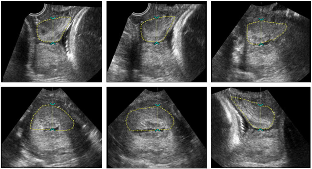
Fig. 1 Six contours of the cervix in 30° increments of rotation with a roller ball cursor. Six anterior cervix contours in 30° rotation increments.
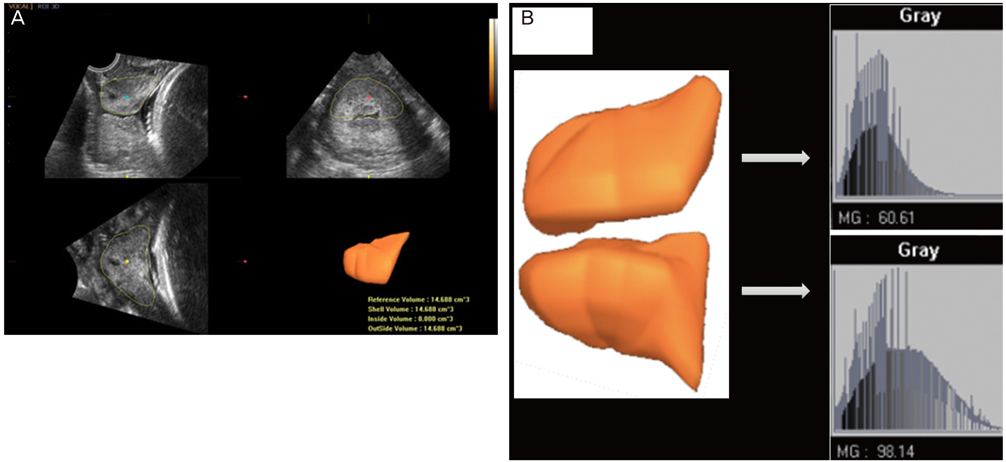
Fig. 2 Three-dimensional ultrasound cervical volume measurement. (A) Multiplanar display of the anterior cervical lip: longitudinal plane in the upper left quadrant, transverse plane in the upper right quadrant and coronal plane in the lower left quadrant. The resultant threedimensional model of the anterior cervix can be seen in the lower right image. (B) Gray-level histogram of each of the lips of the cervix shown at higher magnification. The mean gray-scale histogram was calculated as 32.0, which roughly agreed with the peak gray-level on the histogram. The mean gray-scale histogram represents the echogenicity of the region of interest.
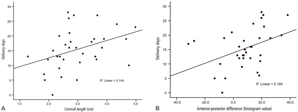
Fig. 3 Correlations between the cervical length and differences of the anterior and posterior cervix with days to delivery using a three-dimensional gray-scale histogram. (A) Cervical length and days to delivery (R = +0.38, P= 0.02, Y=2.931 [x]+6.700). (B) Anterior-posterior histogram difference and delivery day (R = +0.40, P=0.01, Y=0.183 [x]+13.890).
Reference
-
1. Radeka G, Novakov-Mikic A, Ivanovic L. The Bishop score and induction of labor. Med Pregl. 2002; 55:189–194.2. Teixeira C, Lunet N, Rodrigues T, Barros H. The Bishop score as a determinant of labour induction success: a systematic review and meta-analysis. Arch Gynecol Obstet. 2012; 286:739–753.3. Kolkman DG, Verhoeven CJ, Brinkhorst SJ, van der Post JA, Pajkrt E, Opmeer BC, et al. The bishop score as a predictor of labor induction success: a systematic review. Am J Perinatol. 2013; 30:625–630.4. Hendrix NW, Chauhan SP, Morrison JC, Magann EF, Martin JN Jr, Devoe LD. Bishop score: a poor diagnostic test to predict failed induction versus vaginal delivery. South Med J. 1998; 91:248–252.5. Zelig CM, Nichols SF, Dolinsky BM, Hecht MW, Napolitano PG. Interaction between maternal obesity and Bishop score in predicting successful induction of labor in term, nulliparous patients. Am J Perinatol. 2013; 30:75–80.6. Faltin-Traub EF, Boulvain M, Faltin DL, Extermann P, Irion O. Reliability of the Bishop score before labour induction at term. Eur J Obstet Gynecol Reprod Biol. 2004; 112:178–181.7. Crane JM, Hutchens D. Transvaginal sonographic measurement of cervical length to predict preterm birth in asymptomatic women at increased risk: a systematic review. Ultrasound Obstet Gynecol. 2008; 31:579–587.8. Owen J. Evaluation of the cervix by ultrasound for the prediction of preterm birth. Clin Perinatol. 2003; 30:735–755.9. Iams JD, Goldenberg RL, Meis PJ, Mercer BM, Moawad A, Das A, et al. The length of the cervix and the risk of spontaneous premature delivery. National Institute of Child Health and Human Development Maternal Fetal Medicine Unit Network. N Engl J Med. 1996; 334:567–572.10. Owen J, Yost N, Berghella V, Thom E, Swain M, Dildy GA 3rd, et al. Mid-trimester endovaginal sonography in women at high risk for spontaneous preterm birth. JAMA. 2001; 286:1340–1348.11. Podobnik M, Bulic M, Smiljanic N, Bistricki J. Ultrasonography in the detection of cervical incompetency. J Clin Ultrasound. 1988; 16:383–391.12. Barber MA, Medina M, Cabrera F, Romero A, Valle L, Garcia-Hernandez JA. Cervical length vs VOCAL cervical volume for predicting pre-term delivery in asymptomatic women at 20-22 weeks' pregnancy. J Obstet Gynaecol. 2012; 32:648–651.13. Jo YS, Jang DG, Kim N, Kim SJ, Lee G. Comparison of cervical parameters by three-dimensional ultrasound according to parity and previous delivery mode. Int J Med Sci. 2011; 8:673–678.14. Dilek TU, Gurbuz A, Yazici G, Arslan M, Gulhan S, Pata O, et al. Comparison of cervical volume and cervical length to predict preterm delivery by transvaginal ultrasound. Am J Perinatol. 2006; 23:167–172.15. Hoesli IM, Surbek DV, Tercanli S, Holzgreve W. Three dimensional volume measurement of the cervix during pregnancy compared to conventional 2D-sonography. Int J Gynaecol Obstet. 1999; 64:115–119.16. Park IY, Kwon JY, Kwon JY, Hong SC, Choi HM, Kwon HS, et al. Usefulness of cervical volume by three-dimensional ultrasound in identifying the risk for preterm birth. Ultrasound Med Biol. 2011; 37:1039–1045.17. Rozenberg P, Rafii A, Senat MV, Dujardin A, Rapon J, Ville Y. Predictive value of two-dimensional and three-dimensional multiplanar ultrasound evaluation of the cervix in preterm labor. J Matern Fetal Neonatal Med. 2003; 13:237–241.18. Furtado MR, Pires CR, Araujo Junior E, De Souza E, Nardozza LM, Moron AF. Transvaginal grey scale histogram of the cervix at 20-25 weeks of pregnancy. Aust N Z J Obstet Gynaecol. 2010; 50:444–449.19. Kuwata T, Matsubara S, Taniguchi N, Ohkuchi A, Ohkusa T, Suzuki M. A novel method for evaluating uterine cervical consistency using vaginal ultrasound gray-level histogram. J Perinat Med. 2010; 38:491–494.20. Tekesin I, Hellmeyer L, Heller G, Romer A, Kuhnert M, Schmidt S. Evaluation of quantitative ultrasound tissue characterization of the cervix and cervical length in the prediction of premature delivery for patients with spontaneous preterm labor. Am J Obstet Gynecol. 2003; 189:532–539.21. Tekesin I, Wallwiener D, Schmidt S. The value of quantitative ultrasound tissue characterization of the cervix and rapid fetal fibronectin in predicting preterm delivery. J Perinat Med. 2005; 33:383–391.22. Jorn H, Kalf K, Schwann R, Rath W. Grey-scale texture analysis of the cervix in pregnancy. Ultraschall Med. 2006; 27:347–354.23. Rovas L, Sladkevicius P, Strobel E, Valentin L. Reference data representative of normal findings at two-dimensional and three-dimensional gray-scale ultrasound examination of the cervix from 17 to 41 weeks' gestation. Ultrasound Obstet Gynecol. 2006; 27:392–402.24. Rovas L, Sladkevicius P, Strobel E, Valentin L. Intraobserver and interobserver reproducibility of three-dimensional gray-scale and power Doppler ultrasound examinations of the cervix in pregnant women. Ultrasound Obstet Gynecol. 2005; 26:132–137.25. Yilmaz NC, Yigiter AB, Kavak ZN, Durukan B, Gokaslan H. Longitudinal examination of cervical volume and vascularization changes during the antepartum and postpartum period using three-dimensional and power Doppler ultrasound. J Perinat Med. 2010; 38:461–465.26. Rath W, Osmers R, Stuhlsatz HW, Adelmann-Grill BC. Biochemical principles of cervix ripening and dilatation. Z Geburtshilfe Perinatol. 1994; 198:186–195.
- Full Text Links
-
- Actions
-
Cited
- CITED
-
- Close
- Share
-
- Similar articles
-
- Significance of Gray-Level Histogram of the Uterine Cervix as a Predictor of Preterm Birth in Pregnant Women with Preterm Labor
- Evaluation of length, volume and gray-scale histogram of the cervix as predictors of successful induction
- A case of successful normal full term delivery after intramural pregnancy excision in heterotypic intramural pregnancy
- A Case of Uterine Prolapse during Pregnancy
- Transvaginal Ultrasonographic Evaluation of the Uterine Cervix in the Prediction of a Successful Induction of Labor in Term Gestation
