A high resolution genetic mapping of the faded (fe) gene to a region between D10mit156 and D10mit193 on mouse chromosome 10
- Affiliations
-
- 1Department of Medical Genetics, College of Medicine, Hallym University, Chuncheon, Korea. jgsuh@hallym.ac.kr
- 2Institute of Natural Medicine, Hallym University, Chuncheon, Korea.
Abstract
- The C57BL/6J-fe/fe mouse is a coat color mutant. The coat color of the homozygote mouse becomes progressively lighter with advancing age. The faded gene (fe) of C57BL/6J-fe/fe was mapped in a 2.0 cM distal to D10mit191 by our group. To make a high-resolution map, we used the Korean wild mouse (KWHM) for a backcross panel, which was captured in 1995 and has been maintained as an inbred line by our laboratory. In the inter-specific backcross panel (N=400), the fe gene was mapped to 1.0 cM distal to D10mit156. The gene order was defined: centromere -D10mit3/85 (1.3+/-0.6 cM)-D10mit155 (1.3+/-0.6 cM)-D10mit191 (2.0+/-0.7 cM)-D10mit156 (1.0+/-0.5 cM)-fe-D10mit193 (1.3+/-0.6 cM)-D10mit54 (1.0+/-0.5 cM)-D10mit44 (8.5+/-1.4 cM)-D10mit42 (10.0+/-1.5 cM). The measured distance between D10mit191 and D10mit 44 differed in both inter-specific (DBA/2) and intra-specific (KWHM) backcross panels (14.2 vs 13.8 cM). Taken together, our high-resolution linkage map of the fe locus from an intra-specific backcross panel will provide a good entry point to isolate the fe gene.
Keyword
Figure
-
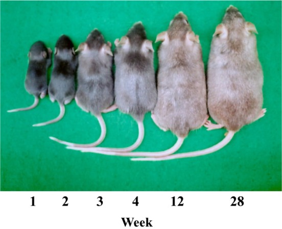
Figure 1 Age-related coat color change in C57BL/6J-fe/fe mice. The coat color of homozygous mice becomes progressively lighter with advancing age (black becoming gray).
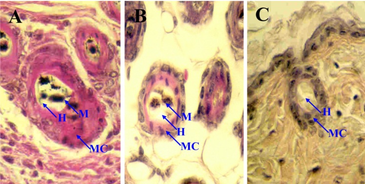
Figure 2 The skin of the C57BL/6J (A) and C57BL/6J-fe/fe mice (B,C). In faded mice, the reduction of melanin content in hair bulbs with age is shown. H&E staining (× 400). M, Melanin; MC, Melanocyte; H, Hair.
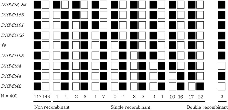
Figure 3 Haplotype analysis of an interspecific backcross between fe and KWHM mice (C57BL/6J-fe/fe X KWHM)F1 X C57BL/6J-fe/fe. Each column represents a chromosomal haplotype identified in the backcross and inherited from the F1 parent. The number of individuals is listed beneath each column. Black boxes represent fe alleles, and white boxs represent KWHM alleles as determined by SSLP scoring with markers indicated at the left.
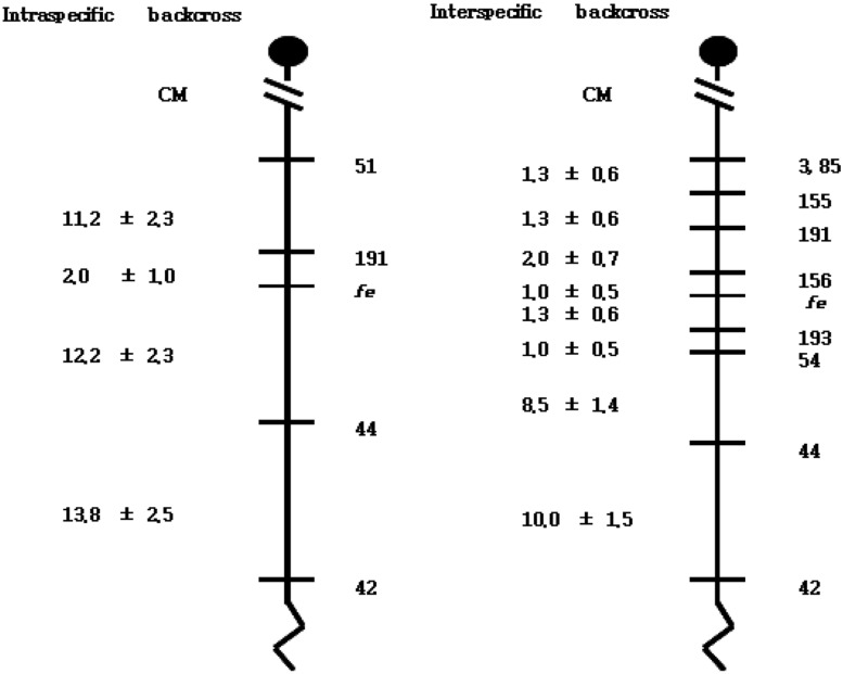
Figure 4 Genetic linkage maps of proximal chromosome 10 as derived from an interspecific backcross panel (C57BL/6J-fe/fe X KWHM)F1 X C57BL/6J-fe/fe) generated in this study. The distances of the intervals between loci and/or markers are not drawn to the same scale, and the locations of mapped human homologs are taken from the Mouse Genome Informatics (The Jackson Laboratory), Distances between intervals are shown in centimorgans±standard error to the left of the map. The linkage map of intra-specific backcross panel came from Oh et al., 2011[21].
Reference
-
1. Steingrímsson E, Copeland NG, Jenkins NA. Mouse coat color mutations: from fancy mice to functional genomics. Dev Dyn. 2006; 235(9):2401–2411. PMID: 16691561.
Article2. Green MC. Genetic variants and strains of the laboratory mouse. 1981. Stuttgart: Gustav Fischer Verlag.3. Marks PW, Bandura JL, Shieh DB, Foernzler D, Beier DR, Kwiatkowski DJ. The spontaneous coat color mutant white nose (wn) maps to murine chromosome 15. Mamm Genome. 1999; 10(7):750–752. PMID: 10384053.4. Kunisada T, Yoshida H, Yamazaki H, Miyamoto A, Hemmi H, Nishimura E, Shultz LD, Nishikawa S, Hayashi S. Transgene expression of steel factor in the basal layer of epidermis promotes survival, proliferation, differentiation and migration of melanocyte precursors. Development. 1998; 125(15):2915–2923. PMID: 9655813.
Article5. O'Reilly FM, Brat DJ, McAlpine BE, Grossniklaus HE, Folpe AL, Arbiser JL. Microphthalmia transcription factor immunohistochemistry: a useful diagnostic marker in the diagnosis and detection of cutaneous melanoma, sentinel lymph node metastases, and extracutaneous melanocytic neoplasms. J Am Acad Dermatol. 2001; 45(3):414–419. PMID: 11511840.6. Lamoreux ML. Strain-specific white-spotting patterns in laboratory mice. Pigment Cell Res. 1999; 12(6):383–390. PMID: 10614578.
Article7. Gariepy CE, Williams SC, Richardson JA, Hammer RE, Yanagisawa M. Transgenic expression of the endothelin-B receptor prevents congenital intestinal aganglionosis in a rat model of Hirschsprung disease. J Clin Invest. 1998; 102(6):1092–1101. PMID: 9739043.
Article8. Richards KA, Fukai K, Oiso N, Paller AS. A novel KIT mutation results in piebaldism with progressive depigmentation. J Am Acad Dermatol. 2001; 44(2):288–292. PMID: 11174389.9. Wang C, Kim E, Attaie A, Smith TN, Wilcox ER, Lalwani AK. A PAX3 polymorphism (T315K) in a family exhibiting Waardenburg Syndrome type 2. Mol Cell Probes. 1998; 12(1):55–57. PMID: 9584079.10. Shanske A, Ferreira JC, Leonard JC, Fuller P, Marion RW. Hirschsprung disease in an infant with a contiguous gene syndrome of chromosome 13. Am J Med Genet. 2001; 102(3):231–236. PMID: 11484199.
Article11. Edery P, Attié T, Amiel J, Pelet A, Eng C, Hofstra RM, Martelli H, Bidaud C, Munnich A, Lyonnet S. Mutation of the endothelin-3 gene in the Waardenburg-Hirschsprung disease (Shah-Waardenburg syndrome). Nat Genet. 1996; 12(4):442–444. PMID: 8630502.
Article12. Tomita Y, Miyamura Y, Kono M, Nakamura R, Matsunaga J. Molecular bases of congenital hypopigmentary disorders in humans and oculocutaneous albinism 1 in Japan. Pigment Cell Res. 2000; 13(Suppl 8):130–134. PMID: 11041370.
Article13. Johnson R, Jackson IJ. Light is a dominant mouse mutation resulting in premature cell death. Nat Genet. 1992; 1(3):226–229. PMID: 1303241.
Article14. Fukuta K, Imamura K, Goto N. Pink-eyed dilution, a coat color mutation in the Japanese field vole (Microtus montebelli). Jikken Dobutsu. 1991; 40(3):375–379. PMID: 1915604.15. Wada A, Okumoto M, Tsudzuki M. Tawny: a novel light coat color mutation found in a wild population of Mus musculus molossinus, a new allele at the melanocortin 1 receptor (Mc1r) locus. Exp Anim. 1999; 48(2):73–78. PMID: 10374067.16. Toyofuku K, Wada I, Valencia JC, Kushimoto T, Ferrans VJ, Hearing VJ. Oculocutaneous albinism types 1 and 3 are ER retention diseases: mutation of tyrosinase or Tyrp1 can affect the processing of both mutant and wild-type proteins. FASEB J. 2001; 15(12):2149–2161. PMID: 11641241.
Article17. Brilliant MH. The mouse p (pink-eyed dilution) and human P genes, oculocutaneous albinism type 2 (OCA2), and melanosomal pH. Pigment Cell Res. 2001; 14(2):86–93. PMID: 11310796.18. Voisey J, Box NF, van Daal A. A polymorphism study of the human Agouti gene and its association with MC1R. Pigment Cell Res. 2001; 14(4):264–267. PMID: 11549109.
Article19. Oh YS, Tomita T. Linkage of faded gene (fe) to chromosome 6 of the mouse. Jikken Dobutsu. 1987; 36(1):73–77. PMID: 3816991.20. Oh YS, Tomita T, Kondo K. Faded, a mutation in the KSB strain of mouse which shows age-related pigment changes. Jikken Dobutsu. 1986; 35(2):131–138. PMID: 3732403.21. Oh SH, Nam Y, Suh JG. Mapping of the Faded (fe) Gene to a Region between D10mit191 and D10mit44 on Mouse Chromosome 10. Lab Anim Res. 2011; 27(1):41–46. PMID: 21826159.22. Suh JG, Yamanishi T, Matsui K, Tanaka K, Wada K. Mapping of the gracile axonal dystrophy (gad) gene to a region between D5Mit197 and D5Mit113 on proximal mouse chromosome 5. Genomics. 1995; 27(3):549–551. PMID: 7558041.
Article23. Green EL. Linkage, recombination and mapping. Genetics and probability in animal breeding experiments. 1981. New York: Oxford University Press;p. 77–133.
Article24. Moriwaki K, Shiroishi T, Yonekawa H. Genetics in wild mice: Its application to biomedical research. 1994. Japan: Japan Scientific societies press;p. 141–295.
- Full Text Links
-
- Actions
-
Cited
- CITED
-
- Close
- Share
-
- Similar articles
-
- Mapping of the Faded (fe) Gene to a Region between D10mit191 and D10mit44 on Mouse Chromosome 10
- Large-scale Genotyping and Genetic Mapping in Plasmodium Parasites
- Characterization of microsatellite markers covering chromosome 1 in the Korean and Japanese populations
- Molecular Genetic Screening for the SRY(Sex Determining Region of the Y chromosme) Gene in Turner Syndrome Patients
- Inner Ear Gene Therapy in Mouse Models of Genetic Hearing Loss
