Retroperitoneoscopic Nephrectomy for a Horseshoe Kidney with Unilateral Severe Hydronephrosis and Ureteral Hypoplasia
- Affiliations
-
- 1Department of Urology, Wonkwang University School of Medicine, Iksan, Korea. uro94c@wonkwang.ac.kr
- 2Department of Pediatrics, Wonkwang University School of Medicine, Iksan, Korea.
Abstract
- A horseshoe kidney is the most common renal fusion anomaly. It is well known that horseshoe kidneys may be associated with many urological problems, including calculi, vesicoureteral reflux, and ureteropelvic junction obstruction. However, a horseshoe kidney with unilateral severe hydronephrosis and ureteral hypoplasia is very rare. We report an 11-year-old female who underwent a retroperitoneoscopic nephrectomy for a horseshoe kidney with severe hydronephrosis and unilateral ureteral hypoplasia.
Keyword
Figure
-
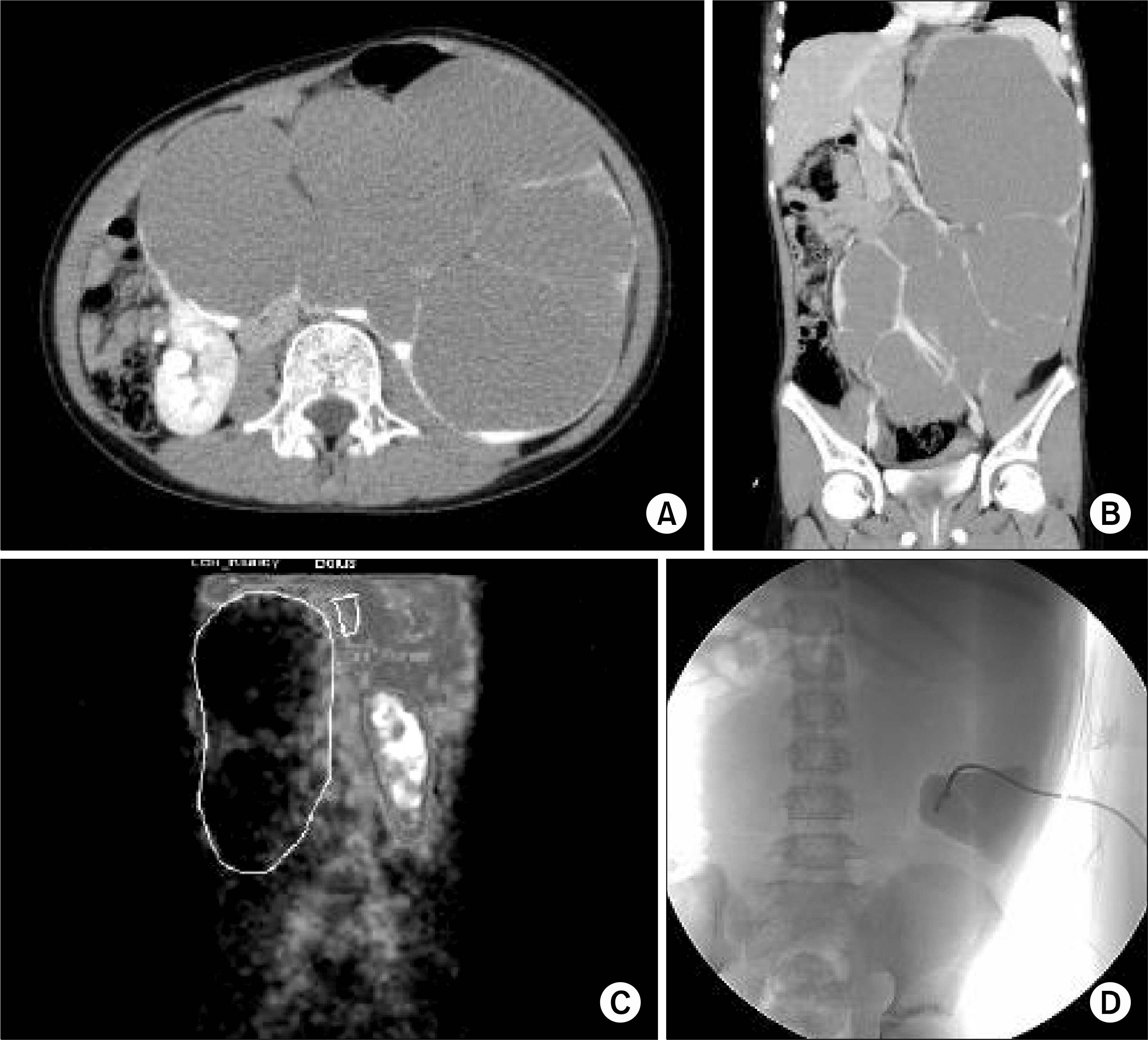
Fig. 1 Imaging studies. (A) and (B) enhanced computed tomography scan, transverse and coronary images, showing a horseshoe kidney with very large hydronephrosis of the left kidney. (C) The 99mTc-MAG3 renal scan showed decreased renal uptake. (D) Insertion of a percutaneous nephrostomy was performed to reduce the size of the hydronephrosis and mark the site so that it could be easily seen during the operation.
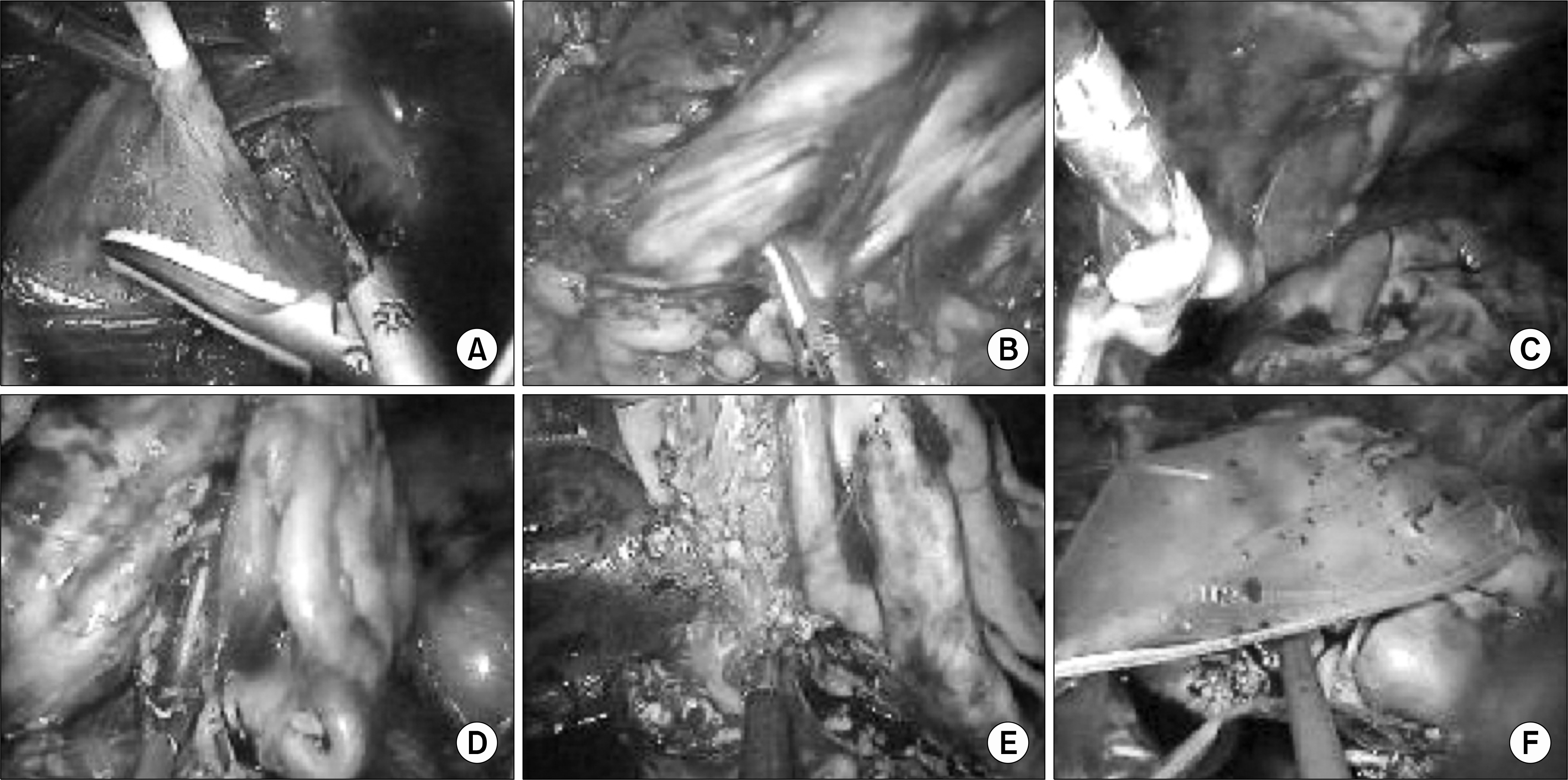
Fig. 2 Intraoperative retroperitoneoscopic nephrectomy procedure for a horseshoe kidney with severe hydronephrosis due to unilateral ureteral hypoplasia. (A) The percutaneous nephrostomy tube was checked. (B) The Gerota's fascia was opened and the kidney was freed. (C) Thread-like hypoplasia of the ureter was seen. (D) Renal arteries and veins were clipped and cut. (E) The isthmus was divided by ultrasonic scissors (SonosurgⓇ). (F) The specimen was put into a LapSacⓇ and extracted.

Fig. 3 Gross findings. (A) Excised kidney showing severe hydronephrosis. (B) Distal ureteral hypoplasia (arrow).
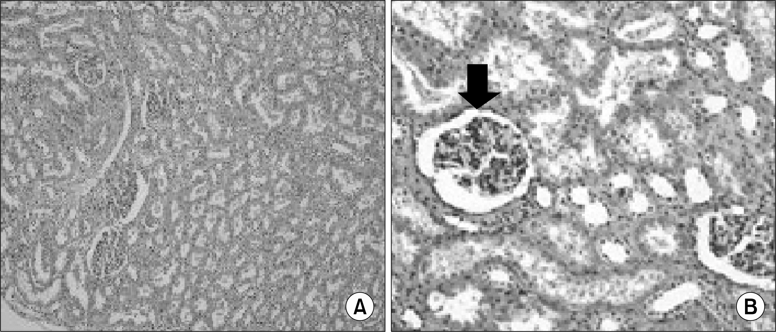
Fig. 4 Pathologic findings. (A) H&E, x100, Nonfunctioning kidney with chronic inflammation. (B) H&E, x200, A few normal nephrons (arrow).
Reference
-
1.Bauer SB. Anomalies of the upper urinary tract. Wein AJ, Kavoussi LR, Novick AC, Partin AW, Peters CA, editors. editors.Campbell-Walsh urology. 9th ed.Philadelphia: Saunders;2007. 3269-304.2.Shimkus EM., Mekhanna I. Hydronephrosis in a horseshoe kidney. Urol Nefrol (Mosk). 1993. 3:48–51.3.Riedl CR., Huebner WA., Schramek P., Pflueger H. Laparo-scopic hemi-nephrectomy in a horseshoe kidney. Br J Urol. 1995. 76:140–1.
Article4.Yohannes P., Smith AD. The endourological management of complications associated with horseshoe kidney. J Urol. 2002. 168:5–8.
Article5.Pearle MS., Nakada SY. Laparoscopic nephrectomy: retroperitoneal approach. Semin Laparosc Surg. 1996. 3:75–83.
Article6.Beck AD. The effect of intra-uterine urinary obstruction upon the development of the fetal kidney. J Urol. 1971. 105:784–9.
Article7.Glick PL., Harrison MR., Noall RA., Villa RL. Correction of congenital hydronephrosis in utero III. Early mid-trimester ureteral obstruction produces renal dysplasia. J Pediatr Surg. 1983. 18:681–7.
Article8.Allen TD., Husmann DA. Ureteropelvic junction obstruction associated with ureteral hypoplasia. J Urol. 1989. 142:353–5.
Article
- Full Text Links
-
- Actions
-
Cited
- CITED
-
- Close
- Share
-
- Similar articles
-
- Three Cases of Horseshoe Kidneys with Complications.
- Horseshoe Kidney associated with Giant Hydronephrosis
- A Case of Horseshoe Kidney Associated with Hydronephrosis and Staghorn Calculi
- Varicose Vein Caused by Giant Hydronephrosis Associated with Horseshoe Kidney
- Horseshoe Kidney Associated with Multiple Renal Stone and Hydronephrosis
