Muscular invasion by oral squamous cell carcinoma of the posterior mandibular alveolar ridge is associated with cervical lymph node metastasis
- Affiliations
-
- 1Department of Oral and Maxillofacial Surgery, School of Dentistry, Seoul National University, Seoul, Korea. myoungh@snu.ac.kr
- KMID: 2309254
- DOI: http://doi.org/10.5125/jkaoms.2016.42.3.133
Abstract
OBJECTIVES
To assess the association between muscle invasion by oral squamous cell carcinoma of the posterior mandibular alveolar ridge and cervical lymph node metastasis on the basis of preoperative magnetic resonance imaging (MRI).
MATERIALS AND METHODS
Twenty-six patients with oral squamous cell carcinoma of the posterior mandibular alveolar ridge were evaluated by MRI. The associations between cervical lymph node metastasis and independent factors evaluated by MRI were analyzed. Overall survival was also analyzed in this manner. Representative biopsy specimens were stained with anti-podoplanin and anti-CD34 antibodies.
RESULTS
Mylohyoid muscle invasion was associated with cervical lymph node metastasis. A combinational factor of mylohyoid and/or buccinator muscle invasion was also associated with cervical lymph node metastasis. Cervical lymph node metastasis and masticator space invasion had a negative effect on overall survival. No lymphatic vessels were identified near the tumor invasion front within the mandible. In contrast, lymphatic vessels were identified near the front of tumor invasion in the muscles.
CONCLUSION
This study demonstrates an association between muscular invasion by oral squamous cell carcinoma of the posterior mandibular alveolar ridge and cervical lymph node metastasis.
Keyword
MeSH Terms
Figure
-
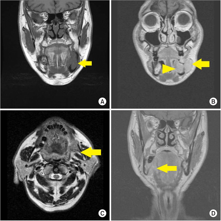
Fig. 1 Anatomical factors assessed on preoperative magnetic resonance imaging. Examples of bone marrow invasion (arrow) (A), sublingual space invasion (arrowhead) and buccinator muscle invasion (arrow) (B), masticator space invasion (arrow) (C), and mylohyoid muscle invasion (arrow) (D) are shown.
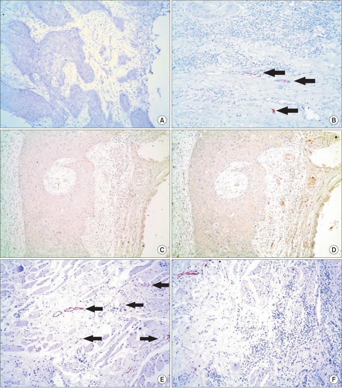
Fig. 2 Immunohistochemical stainings of sections from representative specimens are shown. No lymphatic vessels are identified in the tumor front within the marrow space of the mandible (A), while lymphatic vessels (arrows) are present at the submocosa overlying the mandible (B). Both Fig. 2. A and 2. B are different areas of the same section stained with anti-podoplanin antibody (A: ×100, B: ×200). With no evidence of lymphatic vessels near the bone marrow invasive tumor front in the anti-podoplanin-stained section (C) (×100), the capillary structures are variously stained by anti-CD34 antibody in a serial section (D) (×100). In contrast with the bone marrow, muscles attached to the mandible contain numerous lymphatic vessels (arrows) (E), thus when the tumor invades the muscles, they have close access to the lymphatic vessels (F). Fig. 2. E and 2. F stained with anti-podoplanin antibody (E: ×200, F: ×100).
Reference
-
1. Choi KK, Kim MJ, Yun PY, Lee JH, Moon HS, Lee TR, et al. Independent prognostic factors of 861 cases of oral squamous cell carcinoma in Korean adults. Oral Oncol. 2006; 42:208–217. PMID: 16249114.
Article2. Geum DH, Roh YC, Yoon SY, Kim HG, Lee JH, Song JM, et al. The impact factors on 5-year survival rate in patients operated with oral cancer. J Korean Assoc Oral Maxillofac Surg. 2013; 39:207–216. PMID: 24471047.
Article3. Woolgar JA, Rogers SN, Lowe D, Brown JS, Vaughan ED. Cervical lymph node metastasis in oral cancer: the importance of even microscopic extracapsular spread. Oral Oncol. 2003; 39:130–137. PMID: 12509965.
Article4. Hiratsuka H, Miyakawa A, Nakamori K, Kido Y, Sunakawa H, Kohama G. Multivariate analysis of occult lymph node metastasis as a prognostic indicator for patients with squamous cell carcinoma of the oral cavity. Cancer. 1997; 80:351–356. PMID: 9241067.
Article5. Merritt RM, Williams MF, James TH, Porubsky ES. Detection of cervical metastasis. A meta-analysis comparing computed tomography with physical examination. Arch Otolaryngol Head Neck Surg. 1997; 123:149–152. PMID: 9046281.
Article6. Krestan C, Herneth AM, Formanek M, Czerny C. Modern imaging lymph node staging of the head and neck region. Eur J Radiol. 2006; 58:360–366. PMID: 16687230.
Article7. Veit P, Ruehm S, Kuehl H, Stergar H, Mueller S, Bockisch A, et al. Lymph node staging with dual-modality PET/CT: enhancing the diagnostic accuracy in oncology. Eur J Radiol. 2006; 58:383–389. PMID: 16476533.
Article8. Civantos F, Zitsch R, Bared A. Sentinel node biopsy in oral squamous cell carcinoma. J Surg Oncol. 2007; 96:330–336. PMID: 17879335.
Article9. Yu MG, Ryu SY. Usefulness of 18F-FDG PET/CT in the evaluation of cervical lymph node metastasis in patients with oral cancer. J Korean Assoc Oral Maxillofac Surg. 2009; 35:213–220.10. Kang JH, Ahn KM, Lee SW, Myoung H, Lee JH, Kim MJ. Neck dissection of clinically N0 Neck of oral squamous cell carcinoma & pathologic comparison. J Korean Assoc Oral Maxillofac Surg. 2007; 33:591–596.11. Kalluri R, Weinberg RA. The basics of epithelial-mesenchymal transition. J Clin Invest. 2009; 119:1420–1428. PMID: 19487818.
Article12. Grille SJ, Bellacosa A, Upson J, Klein-Szanto AJ, van Roy F, Lee-Kwon W, et al. The protein kinase Akt induces epithelial mesenchymal transition and promotes enhanced motility and invasiveness of squamous cell carcinoma lines. Cancer Res. 2003; 63:2172–2178. PMID: 12727836.13. Qiao B, Johnson NW, Gao J. Epithelial-mesenchymal transition in oral squamous cell carcinoma triggered by transforming growth factor-beta1 is Snail family-dependent and correlates with matrix metalloproteinase-2 and -9 expressions. Int J Oncol. 2010; 37:663–668. PMID: 20664935.
Article14. Ogura I, Kurabayashi T, Amagasa T, Okada N, Sasaki T. Mandibular bone invasion by gingival carcinoma on dental CT images as an indicator of cervical lymph node metastasis. Dentomaxillofac Radiol. 2002; 31:339–343. PMID: 12424630.
Article15. Breiteneder-Geleff S, Soleiman A, Kowalski H, Horvat R, Amann G, Kriehuber E, et al. Angiosarcomas express mixed endothelial phenotypes of blood and lymphatic capillaries: podoplanin as a specific marker for lymphatic endothelium. Am J Pathol. 1999; 154:385–394. PMID: 10027397.16. Fukano H, Matsuura H, Hasegawa Y, Nakamura S. Depth of invasion as a predictive factor for cervical lymph node metastasis in tongue carcinoma. Head Neck. 1997; 19:205–210. PMID: 9142520.
Article17. Asakage T, Yokose T, Mukai K, Tsugane S, Tsubono Y, Asai M, et al. Tumor thickness predicts cervical metastasis in patients with stage I/II carcinoma of the tongue. Cancer. 1998; 82:1443–1448. PMID: 9554518.
Article18. Ogura I, Amagasa T, Miyakura T. Correlation between tumor consistency and cervical metastasis in tongue carcinoma. Head Neck. 2000; 22:229–233. PMID: 10748445.
Article19. Kurokawa H, Yamashita Y, Takeda S, Zhang M, Fukuyama H, Takahashi T. Risk factors for late cervical lymph node metastases in patients with stage I or II carcinoma of the tongue. Head Neck. 2002; 24:731–736. PMID: 12203797.
Article20. Eicher SA, Overholt SM, el-Naggar AK, Byers RM, Weber RS. Lower gingival carcinoma. Clinical and pathologic determinants of regional metastases. Arch Otolaryngol Head Neck Surg. 1996; 122:634–638. PMID: 8639295.
Article21. Ogura I, Kurabayashi T, Sasaki T, Amagasa T, Okada N, Kaneda T. Maxillary bone invasion by gingival carcinoma as an indicator of cervical metastasis. Dentomaxillofac Radiol. 2003; 32:291–294. PMID: 14709602.
Article22. Edwards JR, Williams K, Kindblom LG, Meis-Kindblom JM, Hogendoorn PC, Hughes D, et al. Lymphatics and bone. Hum Pathol. 2008; 39:49–55. PMID: 17904616.
Article
- Full Text Links
-
- Actions
-
Cited
- CITED
-
- Close
- Share
-
- Similar articles
-
- A clinico-statistical study on cervical lymph node metastasis oforal squamous cell carcinoma
- A case of extremely early cervical adenocarcinoma diagnosed only by endocervical curettage with macroscopic pelvic lymph node metastases
- A Case of Squamous Cell Carcinoma of the Nail Bed with Lymph Node Metastasis
- A CLINICAL STUDY ON CERVICAL LYMPH NODE METASTASIS OF ORAL CANCER
- The Clinicopathological Study On The Relation Of Microvessel Density And Aggressiveness In Oral Squamous Cell Carcinoma
