A dose monitoring system for dental radiography
- Affiliations
-
- 1Department of Oral and Maxillofacial Radiology and Dental Research Institute, School of Dentistry, Seoul National University, Seoul, Korea. raylee@snu.ac.kr
- 2Division of Oral and Maxillofacial Radiology, Department of Basic Science, Faculty of Dentistry, University of Health Sciences, Vientiane, Laos.
- 3Interdisciplinary Program in Radiation, Applied Life Sciences Major, College of Medicine, BK21, and Dental Research Institute, Seoul National University, Seoul, Korea.
- 4School of Computer Science Engineering, Seoul National University, Seoul, Korea.
- KMID: 2308873
- DOI: http://doi.org/10.5624/isd.2016.46.2.103
Abstract
- PURPOSE
The current study investigates the feasibility of a platform for a nationwide dose monitoring system for dental radiography. The essential elements for an unerring system are also assessed.
MATERIALS AND METHODS
An intraoral radiographic machine with 14 X-ray generators and five sensors, 45 panoramic radiographic machines, and 23 cone-beam computed tomography (CBCT) models used in Korean dental clinics were surveyed to investigate the type of dose report. A main server for storing the dose data from each radiographic machine was prepared. The dose report transfer pathways from the radiographic machine to the main sever were constructed. An effective dose calculation method was created based on the machine specifications and the exposure parameters of three intraoral radiographic machines, five panoramic radiographic machines, and four CBCTs. A viewing system was developed for both dentists and patients to view the calculated effective dose. Each procedure and the main server were integrated into one system.
RESULTS
The dose data from each type of radiographic machine was successfully transferred to the main server and converted into an effective dose. The effective dose stored in the main server is automatically connected to a viewing program for dentist and patient access.
CONCLUSION
A patient radiation dose monitoring system is feasible for dental clinics. Future research in cooperation with clinicians, industry, and radiologists is needed to ensure format convertibility for an efficient dose monitoring system to monitor unexpected radiation dose.
Keyword
MeSH Terms
Figure
-
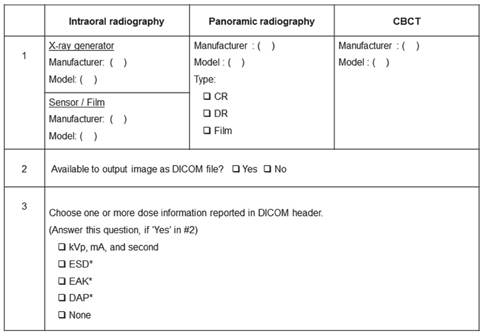
Fig. 1 Items in a questionnaire for dental radiographic machines. *ESD: entrance surface dose, EAK: entrance air kerma, DAP: dose area product.
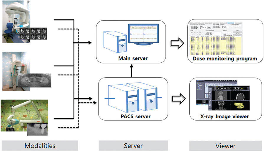
Fig. 2 Flows of the dose report and X-ray image obtained from different radiographic machines to the main server (--- single ouput port, ---- multiple output port).
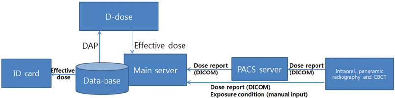
Fig. 3 Schematic view of the dose report transfer, conversion, and store process in the dose monitoring system.
Cited by 1 articles
-
Development of an evidence-based clinical imaging diagnostic guideline for implant planning: Joint recommendations of the Korean Academy of Oral and Maxillofacial Radiology and National Evidence-based Healthcare Collaborating Agency
Min-Ji Kim, Sam-Sun Lee, Miyoung Choi, Eun Ju Ha, Chena Lee, Jo-Eun Kim, Min-Suk Heo
Imaging Sci Dent. 2020;50(1):45-52. doi: 10.5624/isd.2020.50.1.45.
Reference
-
1. United Nations Scientific Committee on the Effects of Atomic Radiation. Sources and effects of ionizing radiation: United Nations Scientific Committee on the Effects of Atomic Radiation: UNSCEAR 2008 report to the General Assembly, with scientific annexes. New York: United Nations;2010.2. American Dental Association. Updated ADA recommendations on dental x-rays. J Can Dent Assoc. 2013; 79:d7.3. Kim HJ. A study on investigation and analysis of medical radiation usage. Final report. Chungcheongbuk-do: Korean Ministry of Food and Drug Safety;2010. 12. Report no.10171Radiology457.4. Roberts JA, Drage NA, Davies J, Thomas DW. Effective dose from cone beam CT examinations in dentistry. Br J Radiol. 2009; 82:35–40.
Article5. Loubele M, Bogaerts R, Van Dijck E, Pauwels R, Vanheusden S, Suetens P, et al. Comparison between effective radiation dose of CBCT and MSCT scanners for dentomaxillofacial applications. Eur J Radiol. 2009; 71:461–468.
Article6. Ludlow JB, Davies-Ludlow LE, Brooks SL, Howerton WB. Dosimetry of 3 CBCT devices for oral and maxillofacial radiology: CB Mercuray, NewTom 3G and i-CAT. Dentomaxillofac Radiol. 2006; 35:219–226.
Article7. Hart D, Hillier MC, Wall BF. National reference doses for common radiographic, fluoroscopic and dental X-ray examinations in the UK. Br J Radiol. 2009; 82:1–12.
Article8. Kim EK. Development of diagnostic reference level in dental x-ray examination in Korea. Fianl report. Chungcheongbuk-do: Korean Ministry of Food and Drug Safety;2009. 11. Report no. 09142Radiology510.9. Kim EK. Evaluation of patient dose for diagnostic reference levels in adult and pediatric dental panoramic radiography. Final report. Chungcheongbuk-do: Korean Ministry of Food and Drug Safety;2014. 07. Report no. 13172Radiology586.10. Technical meeting on patient radition exposure tracking: progress assessment and development of further actions. Conference. In : International Atomic Energy Agency; 2013 Sep; Vienna. Conference code. 44107.11. Baerlocher MO, Talanow R, Baerlocher AF. Radiation passport: an iPhone and iPod touch application to track radiation dose and estimate associated cancer risks. J Am Coll Radiol. 2010; 7:277–280.
Article12. Henriques S. A new way of thinking about patient radiation exposure. Division of public information. In : International Atomic Energy Agency; 2013 Oct; Vienna.13. Rehani MM. Tracking of examination and dose: overview. Radiat Prot Dosimetry. 2015; 165:50–52.
Article14. Rehani MM, Berris T. Radiation exposure tracking: survey of unique patient identification number in 40 countries. AJR Am J Roentgenol. 2013; 200:776–779.
Article15. AlSuwaidi JS, AlMazrouei NK, Pottybindu S, Siraj M, Mathew D, Al Blooshi AA, et al. Patient dose monitoring in Dubai in radiography and interventional procedures. Ann ICRP. 2015; 44:1 Suppl. 249–258.
Article16. Seuri R, Rehani MM, Kortesniemi M. How tracking radiologic procedures and dose helps: experience from Finland. AJR Am J Roentgenol. 2013; 200:771–775.
Article17. Hughes JS, Watson SJ, Jones AL, Oatway WB. Review of the radiation exposure of the UK population. J Radiol Prot. 2005; 25:493–496.
Article18. Hricak H, Brenner DJ, Adelstein SJ, Frush DP, Hall EJ, Howell RW, et al. Managing radiation use in medical imaging: a multifaceted challenge. Radiology. 2011; 258:889–905.
Article
- Full Text Links
-
- Actions
-
Cited
- CITED
-
- Close
- Share
-
- Similar articles
-
- Absorbed and effective dose for periapical radiography using portable and wall type dental X-ray machines
- Skin entrance dose for digital and film radiography in Korean dental schools
- Absorbed and effective dose from periapical radiography by portable intraoral x-ray machine
- Radiography Work Performed by Dental Hygienists according to the Workplace Type
- Leakage and scattered radiation from hand-held dental x-ray unit
