J Rhinol.
2015 May;22(1):59-62. 10.18787/jr.2015.22.1.59.
A Case of Frontal Mococele Treated with Transblepharoplasty Approach Combined with Endoscopic Approach
- Affiliations
-
- 1Department of Otorhinolaryngology-Head and Neck Surgery, Korea University College of Medicine, Seoul, Korea. lhman@korea.ac.kr
- 2Biomedical Sciences, Korea University College of Medicine, Seoul, Korea.
- 3Institute for Medical Devices Clinical Trial Center, Korea University College of Medicine, Seoul, Korea.
- KMID: 2297558
- DOI: http://doi.org/10.18787/jr.2015.22.1.59
Abstract
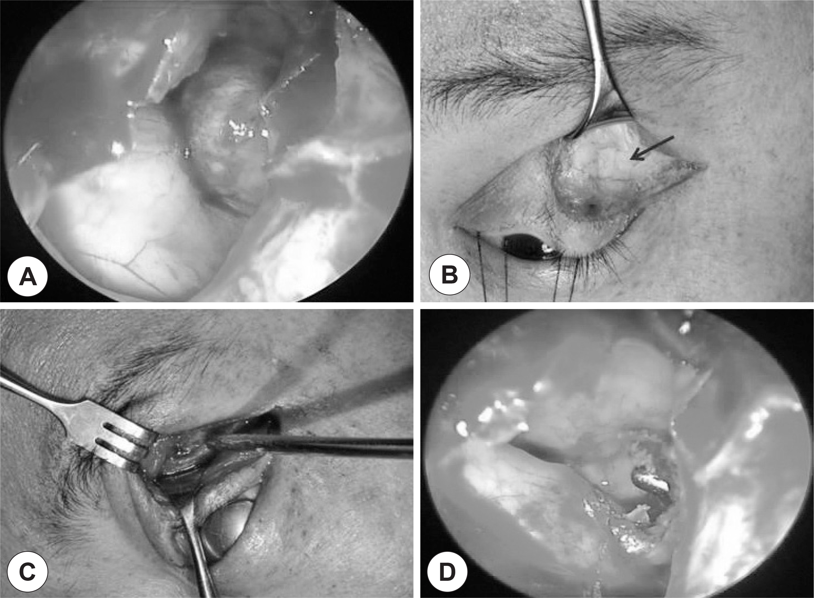
- In recent years, endoscopic sinus marsupialization has become the treatment of choice for the treatment of paranasal sinus mucoceles due to its noninvasiveness and successful outcome. However, mucoceles located at the lateral portion of the frontal sinus and protruding into the orbit with erosion of the frontal sinus floor arestill difficult to address with standard endoscopic sinus surgery techniques. Here, we report a case of a mucocele located atthe lateral side of the frontal sinus and successfully marsupialized with a transblepharoplasty approach combined with an endoscopic approach.
Keyword
MeSH Terms
Figure
Reference
-
References
1). Lund VJ, Milroy CM. Fronto-ethmoidal mucocoeles: a histopathological analysis. J Laryngol Otol. 1991; 105:921–3.
Article2). Constantinidis J, Steinhart H, Schwerdtfeger K, Zenk J, Iro H. Therapy of invasive mucoceles of the frontal sinus. Rhinology. 2001; 39:33–8.3). Kennedy DW, Josephson JS, Zinreich SJ, Mattox DE, Goldsmith MM. Endoscopic sinus surgery for mucoceles: a viable alternative. Laryngoscope. 1989; 99:885–95.4). Bockmuhl U, Kratzsch B, Benda K, Draf W. Surgery for paranasal sinus mucocoeles: efficacy of endonasal micro-endoscopic management and longterm results of 185 patients. Rhinology. 2006; 44:62–7.5). Stumpe MR, Sindwani R, Chandra RK. Endoscopic management of sinus disease with frontal lobe displacement. Am J Rhinol. 2007; 21:324–9.
Article6). Shikowitz MJ, Goldstein MN, Stegnjajic A. Sphenoid sinus mucocele masquerading as a skull base malignancy. Laryngoscope. 1986; 96:1405–10.
Article7). Har-El G. Endoscopic management of 108 sinus mucoceles. Laryngoscope. 2001; 111:2131–4.
Article8). Weber R, Draf W, Kratzsch B, Hosemann W, Schaefer SD. Modern concepts of frontal sinus surgery. Laryngoscope. 2001; 111:137–46.
Article9). Ulualp SO, Carlson TK, Toohill RJ. Osteoplastic flap versus modified endoscopic Lothrop procedure in patients with frontal sinus disease. Am J Rhinol. 2000; 14:21–6.
Article10). Knipe TA, Gandhi PD, Fleming JC, Chandra RK. Transblepharoplasty approach to sequestered disease of the lateral frontal sinus with ophthalmologic manifestations. Am J Rhinol. 2007; 21:100–4.
Article
- Full Text Links
- Actions
-
Cited
- CITED
-
- Close
- Share
- Similar articles
-
- Two Cases of Frontal Sinus Inverted Papilloma Treated With a Combined Bifrontal Craniotomy and Endonasal Endoscopic Approach
- A Case of Endoscopic Sinus Surgery for Lateral Frontal Sinusitis
- Extensive Transbasal Approach to Skull Base Tumor
- A Case of Isolated Frontal Fungal Sinusitis: Treated by Endoscopic Sinus Surgery with Frontal Sinus Minitrephination
- Combination Therapy with Endoscopic Removal and Ambisome for Rhinocerebral Aspergillosis Invading Frontal Lobe: Report of a Case


