MRI Features of Axial Spondyloarthritis and Differential Diagnosis: Focusing on the Spine and Sacroiliac Joint
- Affiliations
-
- 1Department of Radiology, Hanyang University College of Medicine, Seoul, Korea. radsh@hanyang.ac.kr
- KMID: 2297522
- DOI: http://doi.org/10.4078/jrd.2014.21.3.110
Abstract
- The spine and sacroiliac joint are involved in most cases of axial spondyloarthritis. Several pathologic findings from a radiography involving the spine and sacroiliac joint are the diagnostic hallmarks of axial spondyloarthritis. However, these radiographic changes reflect structural damage rather than active inflammation, which may delay diagnosis by several years. Nowadays, the Assessment of SpondyloArthritis international Society (ASAS) has focused on the reassessment of existing classification criteria and the development and validation of diagnostic tools to facilitate early diagnosis and assessment of treatment response. Magnetic resonance (MR) findings are the most remarkable changes with respect to the previously established classification criteria. Familiarity with typical MRI findings of axial spondyloarthritis and differential diseases is important in order to correctly interpret the findings and avoid misdiagnosis.
MeSH Terms
Figure
-

Figure 1. ASAS classification criteria for axial spondyloarthritis (2009) in patients with back pain ≥3 months and age at onset >45 years. ASAS: Assessment of Spondyloarthritis International Society.
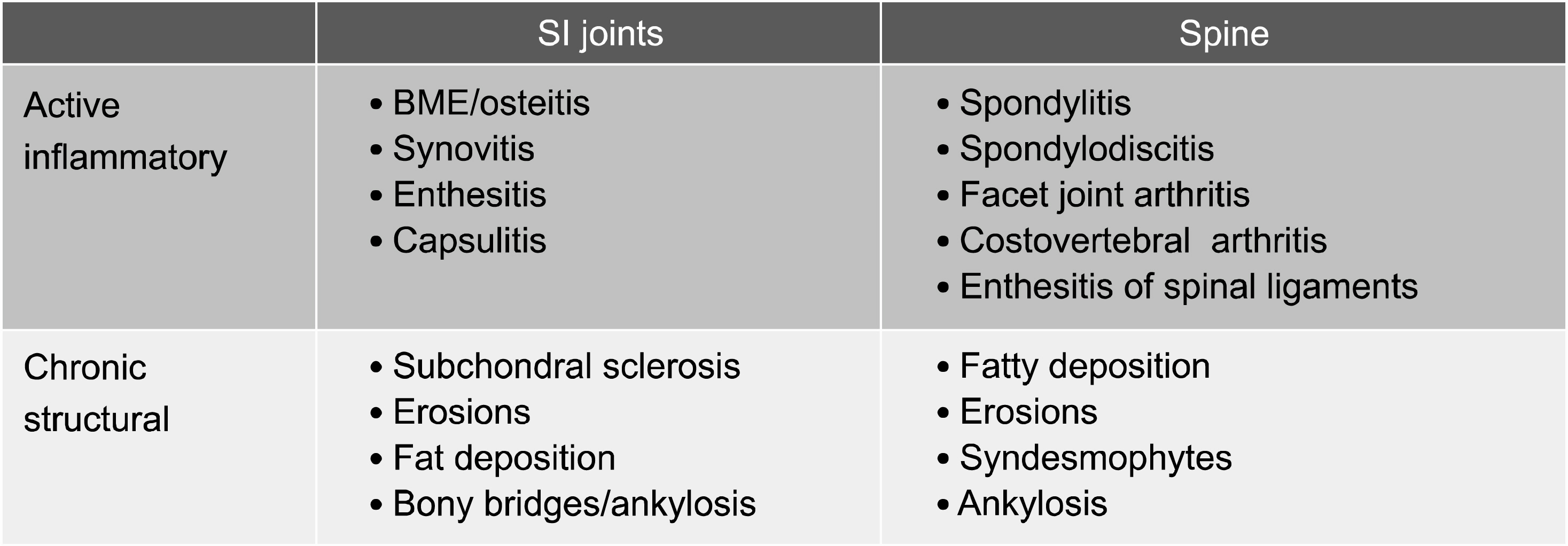
Figure 2. MRI changes in axial spondyloarthritis – ASAS/OMERACT consensual approach. ASAS: Assessment of Spondyloarthritis Inter-national Society, OMERACT: Outcome Measures in Rheumatology.

Figure 3. Non-radiographic axial spondyloarthritis. (A) There is no evidence of sacroiliitis on plain radiography. (B) Hyperintense signal intensity on STIR image and (C) focal enhancement on fat-suppressed Gd-enhanced T1-weighted image in the left sacroiliac joint (arrows) reflect active inflammation. STIR: Short tau inversion recovery.

Figure 4. (A) Oblique coronal T1-weighted, (B) STIR, and (C) fat-suppressed Gd-enhanced T1-weighted MR images of sacroiliac joints show focal bony erosion, subchondral sclerosis and synovitis (arrows).

Figure 5. (A) Oblique coronal T1-weighted, (B) fat-suppressed T2-weighted, and (C) fat-suppressed Gd-enhanced T1-weighted MR images of sacroiliac joints show focal capsulitis in the anterior aspect of left sacroiliac joint (arrows) and active inflammation in the sacrum near left sacroiliac joint.
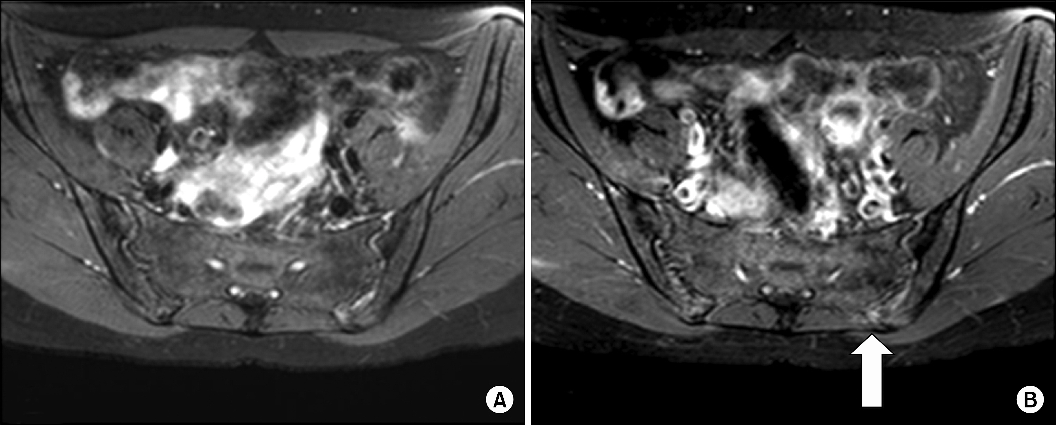
Figure 6. (A) Axial STIR and (B) fat-suppressed Gd-enhanced T1-weighted MR images of sacroiliac joints show enthesitis in the left posterosuperior sacroiliac joint (arrows). STIR: Short tau inversion recovery.
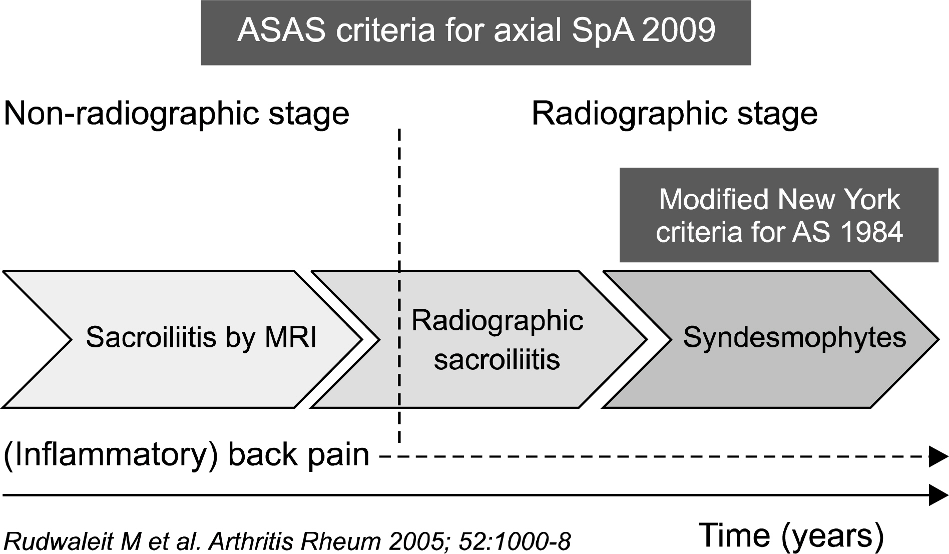
Figure 7. Staging of axial spondyloarthritis.
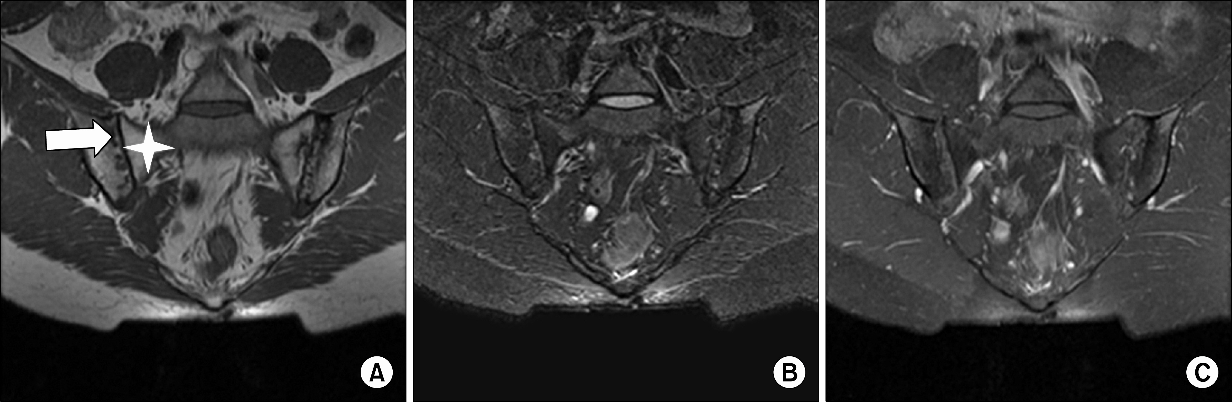
Figure 8. (A) Oblique coronal T1-weighted, (B) fat-suppressed T2-weighted, and (C) fat-suppressed Gd-enhanced T1-weighted MR images of sacroiliac joints show subchondral bone erosions (arrow) and fat deposition (star) around both sacroiliac joints.
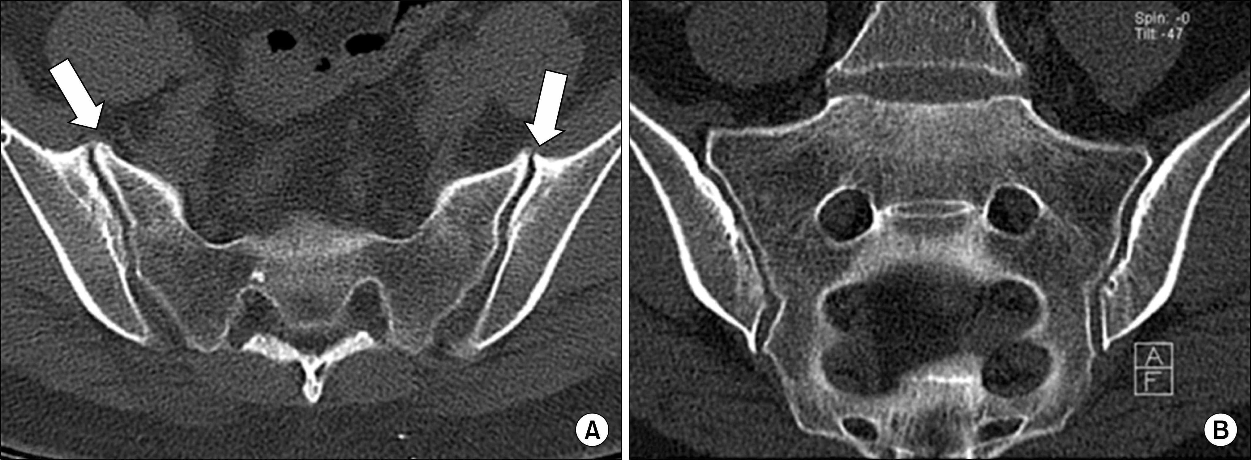
Figure 9. 50-year-old women with degenerative sacroiliitis. (A) Axial and (B) oblique coronal CT scans show osteophytes (arrows) and subchondral sclerosis in the anterior aspect of both sacroiliac joints without definite bony erosion in the both sacroiliac joint.

Figure 10. (A) Plain radiography and (B) oblique coronal CT scans of sacroiliac joint show well-defined trangular sclerosis involving both sacroiliac joints without definite bony erosions. (C) Oblique coronal T1-weighted and (D) fat-suppressed T2-weighted MR images of sacroiliac joints show well-defined low signal intensity in the both ilium near both sacroiliac joints without definite bony erosions characterizing sclerosis.
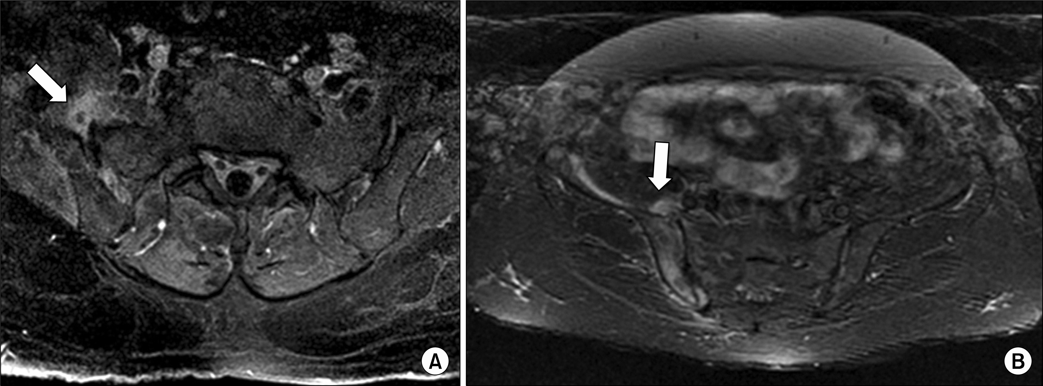
Figure 11. 63-year-old women with right pyogenic sacroiliitis. (A, B) Fat-suppressed Gd-enhanced T1-weighted MR images show focal loculated fluid collection and soft tissue inflammation (arrows) in the anterior aspect of right sacroiliac joint with marrow enhancement in the right ilium characterizing osteomyelitis.

Figure 12. Pelvic abnormalities in DISH. (A) Note the irregular bony excrescences above the both anterior inferior iliac spines (arrow heads) and heterotopic ossification along the right sacrotuberous ligament (empyt arrow) in relation to DISH. (B) The osseous bridges (arrows) extend across the both sacroiliac joints without definite sacroiliitis. DISH: diffuse idiopathic skeletal hyperostosis.
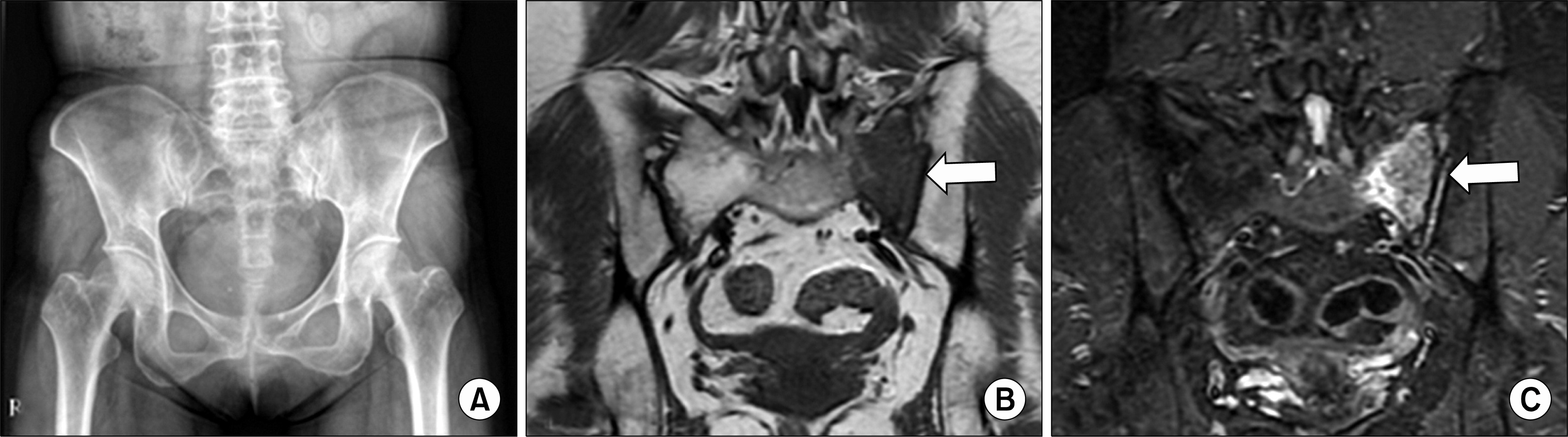
Figure 13. (A) There is no evidence of sacroiliitis on plain radiography of pelvis. Subtle sclerosis in the left sacral wing is suspected. (B) Hypointense signal intensity on T1-weighted image and (C) diffuse edema on STIR in the left sacral wing (arrows) reflect insufficient fracture. STIR: Short tau inversion recovery.
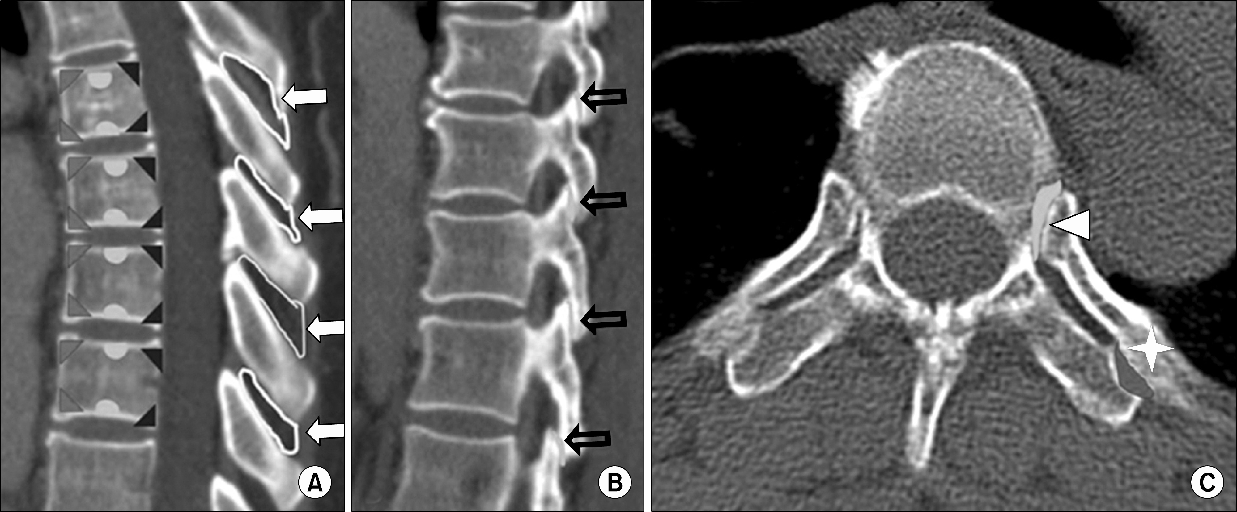
Figure 14. (A, B) Sagittal and (C) axial CT scans of thoracic spine. Spondyloarthritis involving the spine show-vertebral body (triangles), discovertebral junction (semicircles), the interspinous spaces (arrows), facet joints (empty arrows), costovertebral joint (arrow heads), costotransverse joint (asterisks).

Figure 15. (A) Sagittal fat-supp-ressed T2-weighted and (B) fat-su-ppressed Gd-enhanced T1-weighted MR images of lumbar spine show active inflammation in the corners of L4 vertebral bodies (arrows) in spondyloarthritis. (C) Sagittal T1-weighted and (D) fat-suppressed Gd-enhanced T1-weighted MR images of lumbar spine show fat deposition in the corners of L4 vertebral bodies (arrow heads) characterizing inactive lesions.
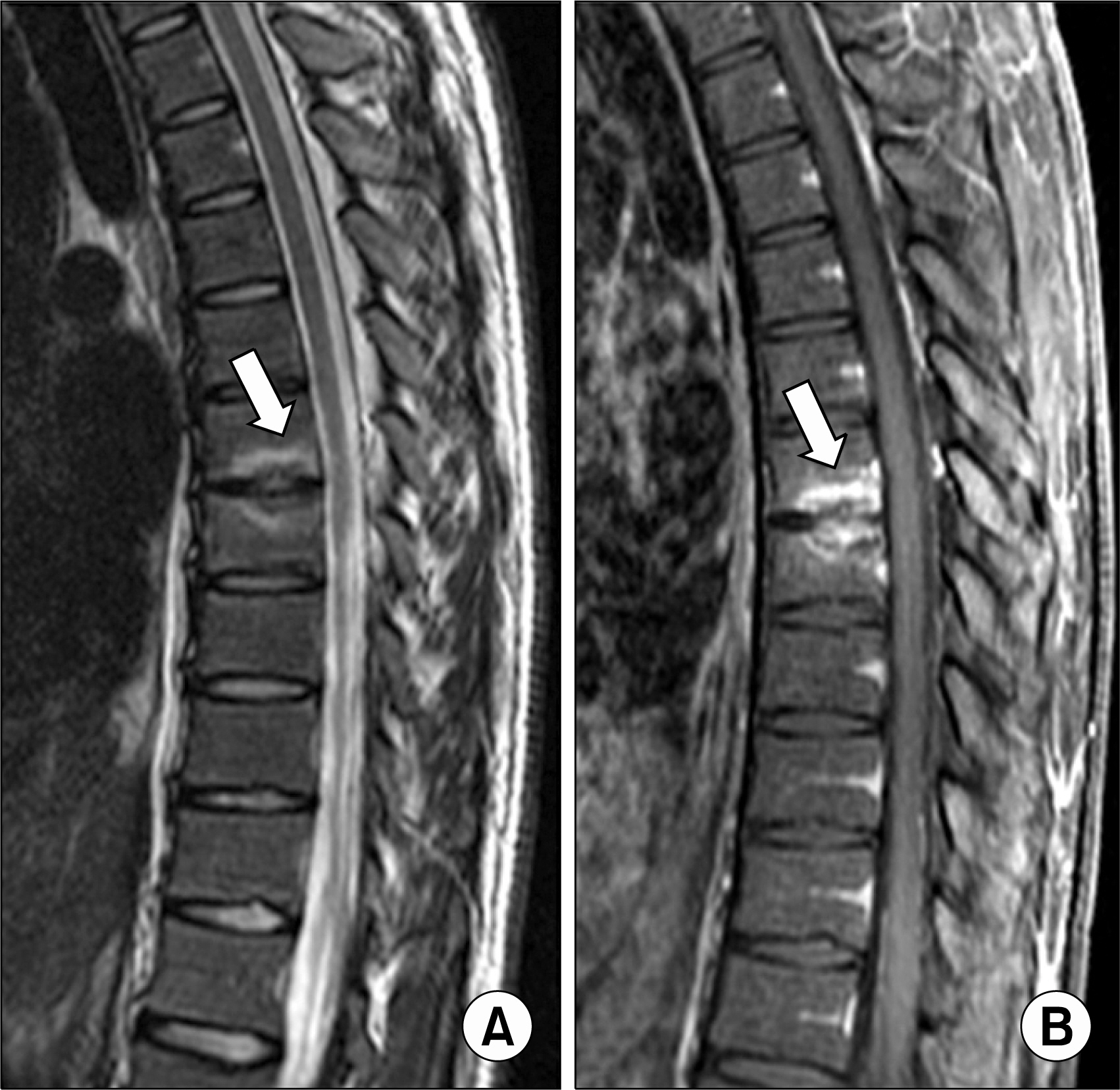
Figure 16. (A) Sagittal fat-suppressed T2-weighted and (B) fat-suppressed Gd-enhanced T1-weighted MR images of thoracic spine show endplate erosion with subchondral marrow edema and inflammation characterizing spondylodiskitis (arrows).
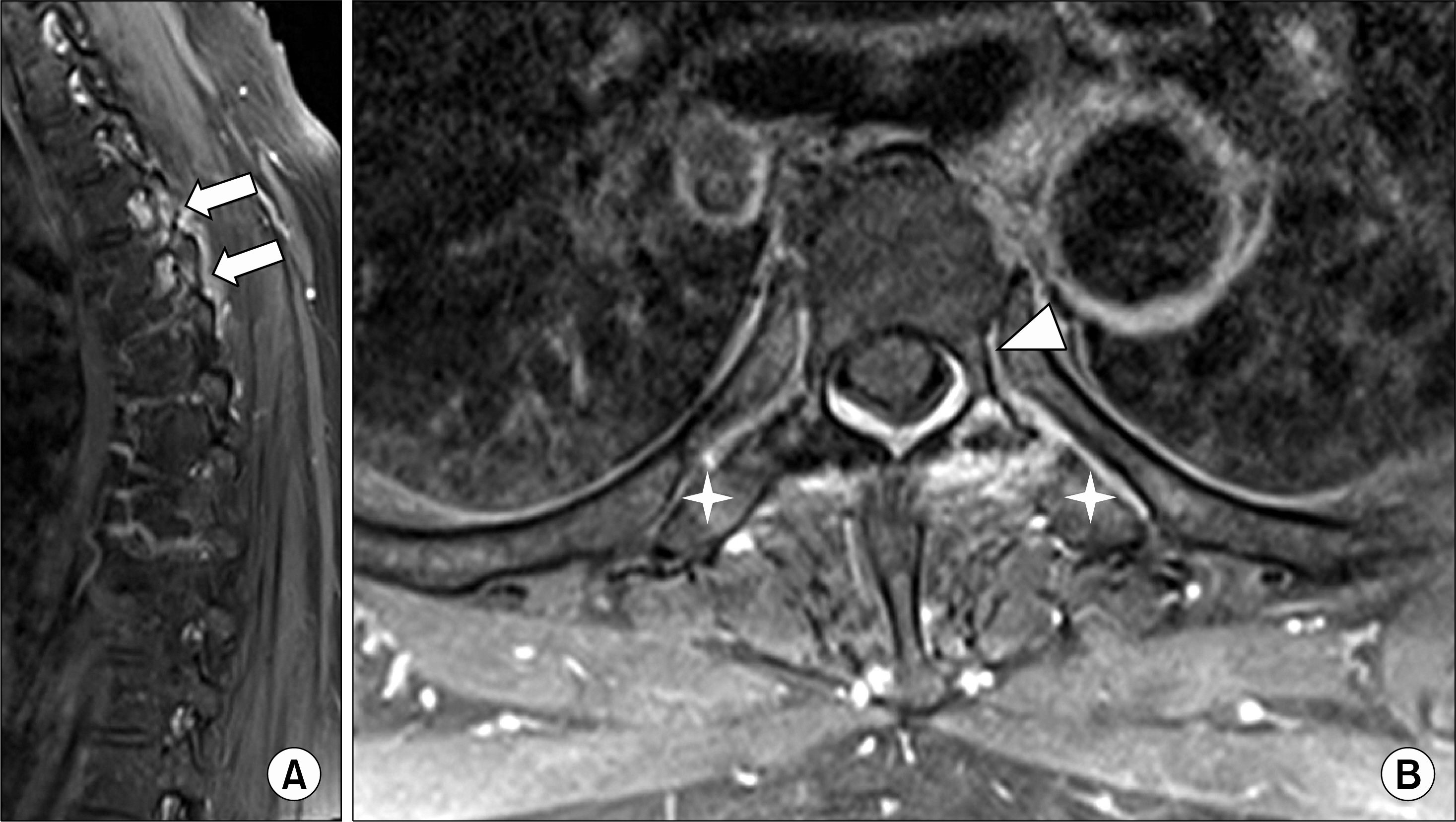
Figure 17. (A) Sagittal and (B) axial fat-suppressed Gd-enhanced T1-weighted MR images of thoracic spine show enhancement in the facet joint (arrows), costovertebral joint (arrow head) and costotransverse joints (asterisks) characterizing active inflammation.

Figure 18. (A) Enthesitis in the L1-2 interspinous and supraspinous ligaments (arrow) on sagittal fat-suppressed Gd-enhanced T1-weighted MR image. (B) AP radiographs of the lumbar spine reveals ossification of the interspinous and supraspinous ligaments, which is producing a vertical central radiodense shadow, the dagger sign.
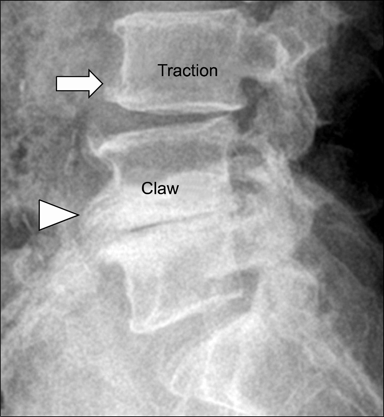
Figure 19. Segmental instability of lumbar spine. A traction osteophyte (arrow) in L3 body develops 2 to 3mm from the edge of the intervertebral disc and horizontal direction. A claw osteophyte (arrow head) in L4 body develops closer to the disc margin and has a sweeping configuration.

Figure 20. (A) Plain radiography of lateral cervical spine, (B) sagittal CT scan and (C) sagittal T1-weighted MR image of cervial spine show extensive anterior bone formation (arrows) and anterior disc extension(empty arrows) without facet involvement (arrow head).
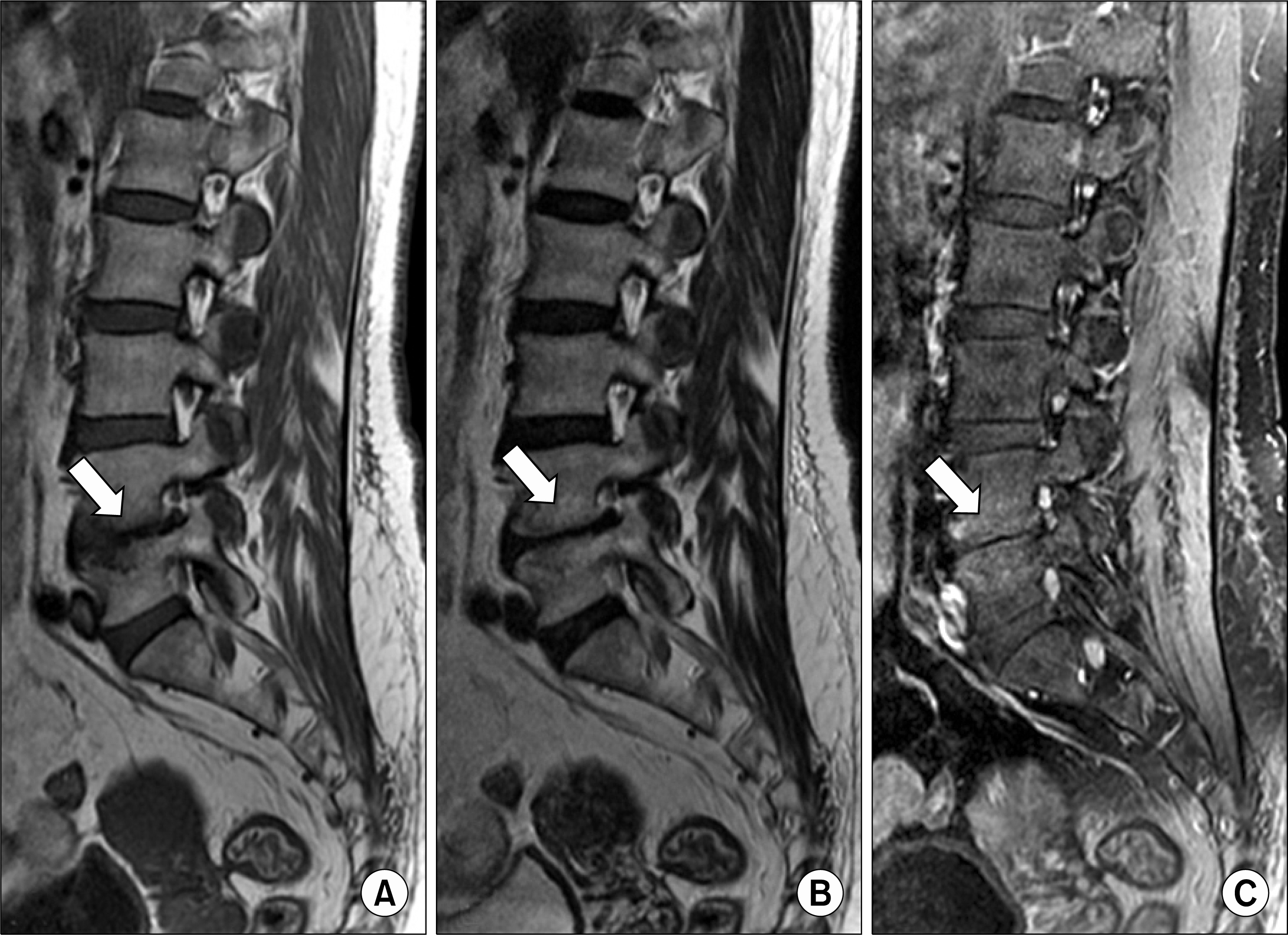
Figure 21. Abnormal intervertebral disc: Modic type I change. (A) Sagittal T1-weighted, (B) T2-weighted and (C) fat-suppressed Gd-enhanced T1-weighted MR images show marrow edema and inflammation without erosion around L4-5 intervertebral disc (arrows) characterizing fibrovascular marrow.
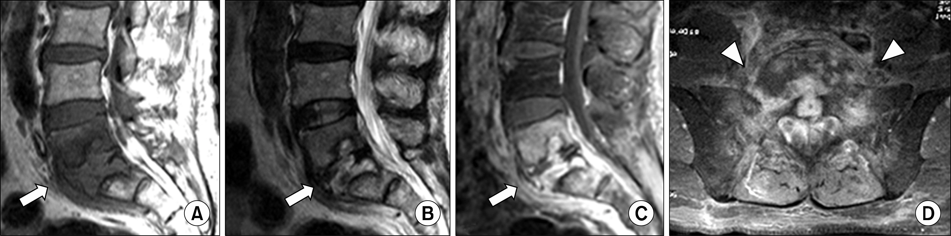
Figure 22. Pyogenic spondylodiskitis involving L5-S1. (A) Sagittal T1-weighted, (B) sagittal T2-weighted, (C) sagittal and (D) axial fat-suppressed Gd-enhanced T1-weighted MR images reveal prominent marrow signal change with end plate erosions (arrows) and paraspinal soft tissue phlegmon (arrow heads) around L5-S1 disc.
Cited by 1 articles
-
Utility of Magnetic Resonance Imaging and Positron Emission Tomography in Rheumatic Diseases
Eun Hye Park, Chong-Hyeon Yoon, Eun Ha Kang, Han Joo Baek
J Rheum Dis. 2020;27(3):136-151. doi: 10.4078/jrd.2020.27.3..
Reference
-
1. van der Linden S, Valkenburg HA, Cats A. Evaluation of diagnostic criteria for ankylosing spondylitis. A proposal for modification of the New York criteria. Arthritis Rheum. 1984; 27:361–8.2. Dougados M, van der Linden S, Juhlin R, Huitfeldt B, Amor B, Calin A, et al. The European Spondylarthropathy Study Group preliminary criteria for the classification of spondylarthropathy. Arthritis Rheum. 1991; 34:1218–27.
Article3. Resnick D, Kransdorf MJ. Bone and joint imaging. 3rd ed.p. 267. Philadelphia: Elsevier Saunders;2005.4. Yochum TR, Rowe LJ. Essentials of skeletal radiology. 2nd ed.p. 877. Baltimore: Williams and Wilkins;1996.5. Braun J, Bollow M, Sieper J. Radiologic diagnosis and pathology of the spondyloarthropathies. Rheum Dis Clin North Am. 1998; 24:697–735.
Article6. Jacobson JA, Girish G, Jiang Y, Resnick D. Radiographic evaluation of arthritis: inflammatory conditions. Radiology. 2008; 248:378–89.
Article7. Rudwaleit M, van der Heijde D, Landewé R, Listing J, Akkoc N, Brandt J, et al. The development of Assessment of SpondyloArthritis international Society classification criteria for axial spondyloarthritis (part II): validation and final selection. Ann Rheum Dis. 2009; 68:777–83.
Article8. Jee WH, McCauley TR, Lee SH, Kim SH, Im SA, Ha KY. Sacroiliitis in patients with ankylosing spondylitis: association of MR findings with disease activity. Magn Reson Imaging. 2004; 22:245–50.
Article9. Rudwaleit M, Jurik AG, Hermann KG, Landewé R, van der Heijde D, Baraliakos X, et al. Defining active sacroiliitis on magnetic resonance imaging (MRI) for classification of axial spondyloarthritis: a consensual approach by the ASAS/OMERACT MRI group. Ann Rheum Dis. 2009; 68:1520–7.
Article10. Bredella MA, Steinbach LS, Morgan S, Ward M, Davis JC. MRI of the sacroiliac joints in patients with moderate to severe ankylosing spondylitis. AJR Am J Roentgenol. 2006; 187:1420–6.
Article11. Lacout A, Rousselin B, Pelage JP. CT and MRI of spine and sacroiliac involvement in spondyloarthropathy. AJR Am J Roentgenol. 2008; 191:1016–23.
Article12. Sieper J, Rudwaleit M, Baraliakos X, Brandt J, Braun J, Burgos-Vargas R, et al. The Assessment of SpondyloArthritis international Society (ASAS) handbook: a guide to assess spondyloarthritis. Ann Rheum Dis. 2009; 68(Suppl 2):ii1–44.
Article13. Jacobson JA, Girish G, Jiang Y, Sabb BJ. Radiographic evaluation of arthritis: degenerative joint disease and variations. Radiology. 2008; 248:737–47.
Article14. Kang HS, Hong SH, Chang HK. Musculoskeletal Radiology. 1st ed.p. 121. Seoul, Korea: Medicalplus;2013.15. Canella C, Schau B, Ribeiro E, Sbaffi B, Marchiori E. MRI in seronegative spondyloarthritis: imaging features and differential diagnosis in the spine and sacroiliac joints. AJR Am J Roentgenol. 2013; 200:149–57.
Article16. Olivieri I, Ferri S, Barozzi L. Osteitis condensans ilii. Br J Rheumatol. 1996; 35:295–7.17. Resnick D, Niwayama G. Osteoporosis. Resnick D, Niwayama G, editors. Diagnosis of bone and joint disorders. 3rd ed.p. 1839. Philadelphia: WB Saunders;1995.18. Modic MT, Steinberg PM, Ross JS, Masaryk TJ, Carter JR. Degenerative disk disease: assessment of changes in vertebral body marrow with MR imaging. Radiology. 1988; 166:193–9.
Article19. Resnick D, Niwayama G. Ankylosing spondylitis. Resnick D, editor. Diagnosis of bone and joint disorders. 3rd ed.p. 1008. Philadelphia: WB Saunders;1995.
Article
- Full Text Links
-
- Actions
-
Cited
- CITED
-
- Close
- Share
-
- Similar articles
-
- Piriformis syndrome as an overlooked cause of pain in a patient with axial spondyloarthritis: a case report
- Ewing's Sarcoma of the Sacroiliac Joint Presenting as Tubercular Sacroiliitis: A Diagnostic Dilemma
- Relief of Symptoms by Early Administration of Infliximab in Patients with Suspected Spondyloarthritis: A Case Report
- A Case of the Lumbar Spine Involvement and Sacroiliitis in a Patient with Gout
- Pyogenic Sacroiliac Joint Infection
