A Case of Cavernous Sinus Dural Arteriovenous Fistula Following Tolosa-Hunt Syndrome
- Affiliations
-
- 1Department of Ophthalmology, Ajou University School of Medicine, Suwon, Korea. mingming8@naver.com
- KMID: 2290502
- DOI: http://doi.org/10.3341/jkos.2016.57.6.977
Abstract
- PURPOSE
To report a case of cavernous sinus dural arteriovenous fistula following Tolosa-Hunt syndrome.
CASE SUMMARY
A 64-year-old female with a history of hypertension, presented with blepharoptosis and periorbital pain in the right eye and diplopia. Her right pupil was dilated. She had right exotropia and right hypertropia with inability to elevate, depress, and adduct the right eye. Magnetic resonance imaging including angiography, revealed hyperintensities in the right cavernous sinus consistent with inflammation and no vascular abnormalities. Three days after oral corticosteroid therapy, the pain disappeared. A presumptive diagnosis was Tolosa-Hunt syndrome presenting as a form of complete oculomotor nerve palsy. Two months later, she experienced severe pain in the right periocular area, even though blepharoptosis was resolved and ocular motility was improved. A rapid response to high-dose intravenous corticosteroids was consistent with recurrence of Tolosa-Hunt syndrome. Three months later, she had normal ocular motility, but developed a conjunctival injection, pulsatile orbital bruits, and exophthalmos in the right eye. Cerebral angiography showed a cavernous sinus dural arteriovenous fistula. She received a transvenous coil embolization and her symptoms markedly improved over 2 months.
CONCLUSIONS
Tolosa-Hunt syndrome, a granulomatous inflammation in the cavernous sinus may be followed by cavernous sinus dural arteriovenous fistula and should be considered during follow-up.
MeSH Terms
-
Adrenal Cortex Hormones
Angiography
Blepharoptosis
Cavernous Sinus*
Central Nervous System Vascular Malformations*
Cerebral Angiography
Diagnosis
Diplopia
Embolization, Therapeutic
Exophthalmos
Exotropia
Female
Follow-Up Studies
Humans
Hypertension
Inflammation
Magnetic Resonance Imaging
Middle Aged
Oculomotor Nerve Diseases
Orbit
Pupil
Recurrence
Strabismus
Tolosa-Hunt Syndrome*
Adrenal Cortex Hormones
Figure
-
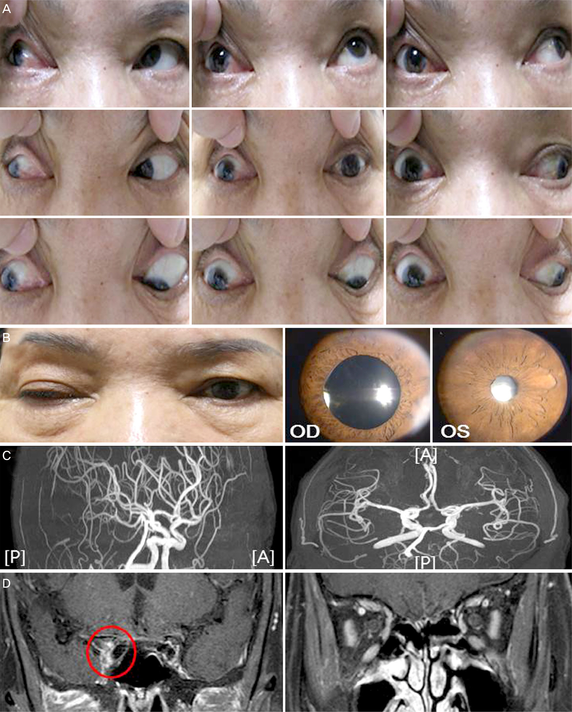
Figure 1. Clinical findings and imaging at initial presentation. (A) Gaze photographs showing severe limitation on supraduction, adduction, and infraduction in the right eye. (B) Photographs showing blepharoptosis, and dilated pupil in the right eye. (C) Brain magnetic resonance angiography (MRA) not showing any abnormality such as aneurysmal dilatation or compressed lesion. (D) Gadolinium enhanced T1-weighted brain magnetic resonance image (MRI) showing increased signal intensity in the right cavernous sinus, indicated by red circle. P = posterior aspect; A = anterior aspect.
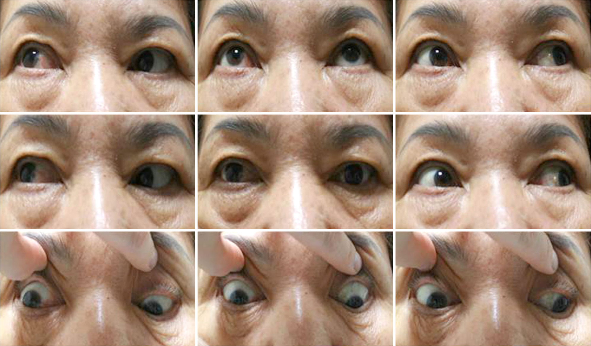
Figure 2. Gaze photographs 2 months after initial presentation when she suffered from severe periocular pain and headache, showing resolution of blepharoptosis and improvement in ocular motility in the right eye.
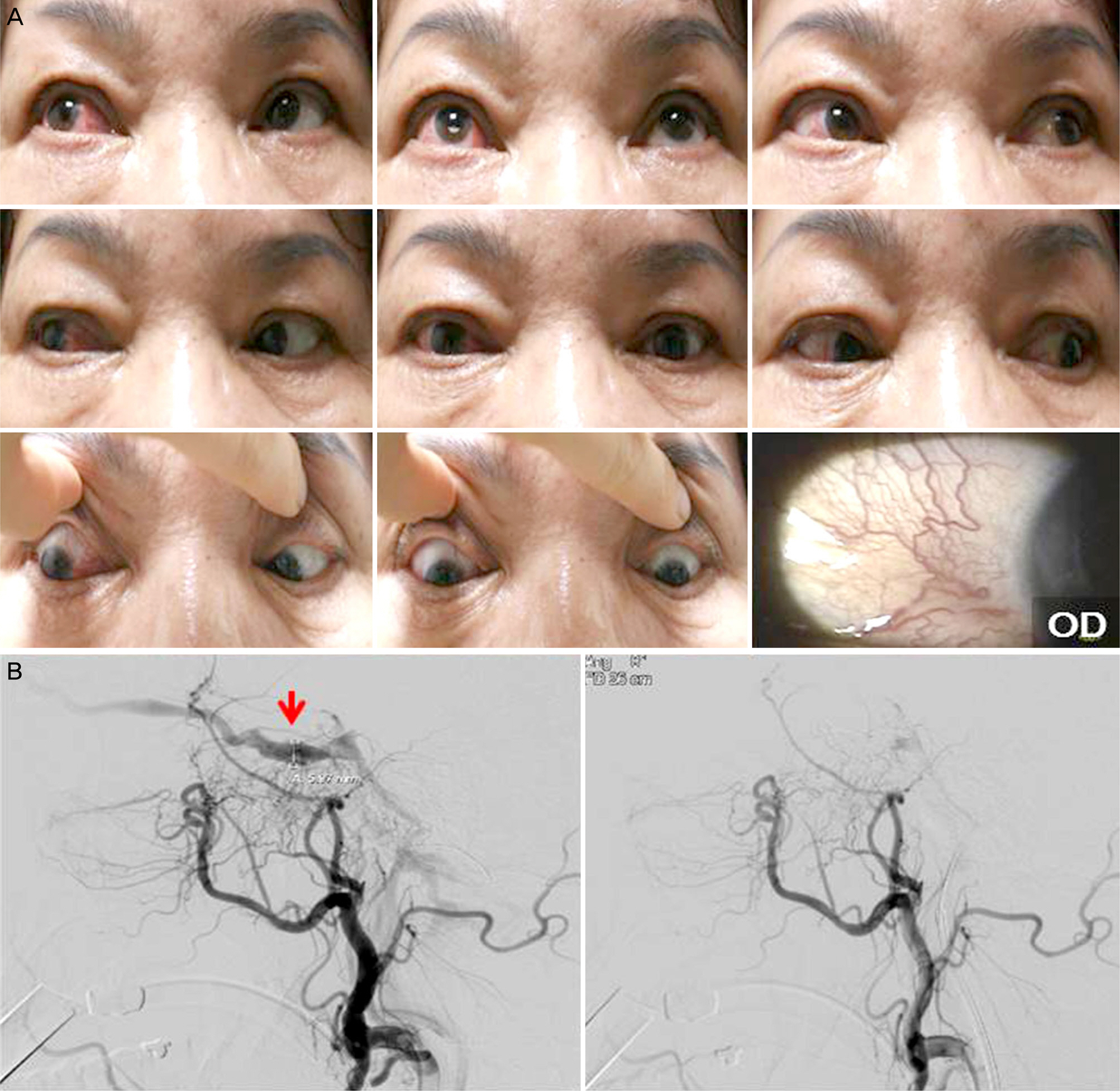
Figure 3. Clinical findings and imaging 5 months after initial presentation. (A) Gaze photographs showing resolution of the right oculomotor nerve palsy, but severe redness in the right eye. Slit lamp photographs showing engorged vessels on the conjunctiva of right eye. (B) Cerebral angiography showing arteriovenous shunt from the right middle meningeal artery and ascending pharyngeal artery, supplied from the right external carotid artery to the cavernous sinus (type C in Barrow classification,10 red arrow, left column), and no more shunt after coil embolization (right column).
Reference
-
References
1. Headache Classification Committee of the International Headache Society (IHS). The international classification of headache abdominal, 3rd edition (beta version). Cephalalgia. 2013; 33:629–808.2. Lee HK, Lee SG. Clinical observations on Tolosa-Hunt syndrome. J Korean Ophthalmol Soc. 2009; 50:1717–23.
Article3. Kim I, Yang S, Lee H, et al. Cavernous sinus dural arteriovenous fistula mimicking Tolosa-Hunt syndrome. J Korean Neurol Assoc. 2014; 32:218–21.4. Ahn JH, Jung JH, Choi KD, Choi HY. The clinical characteristics and endovascular management outcomes of dural carotid abdominal fistulas. J Korean Ophthalmol Soc. 2011; 52:332–7.5. Kim C, Khwarg SI, Han MH. The clinical characteristics and treatment outcomes of dural carotid-cavernous sinus fistula. J Korean Ophthalmol Soc. 2003; 44:1463–74.6. Jang JW, Lee SY, Kim SJ. Clinical characteristics of dural abdominal sinus fistula. J Korean Ophthalmol Soc. 2000; 41:737–43.7. Itokawa K, Fukui M, Yamamoto T, et al. Dural arteriovenous abdominal as a possible cause of Tolosa-Hunt syndrome: a case report. J Neurol. 2010; 257:846–7.8. Sugano H, Iizuka Y, Arai H, Sato K. Progression of Tolosa-Hunt syndrome to a cavernous dural arteriovenous fistula: a case report. Headache. 2003; 43:122–6.
Article9. Brazis PW, Capobianco DJ, Chang FL, et al. Low flow dural abdominal shunt: another cause of “sinister” Tolosa-Hunt syndrome. Headache. 1994; 34:523–5.10. Barrow DL, Spector RH, Braun IF, et al. Classification and abdominal of spontaneous carotid-cavernous sinus fistulas. J Neurosurg. 1985; 62:248–56.11. Houser OW, Campbell JK, Campbell RJ, Sundt TM Jr. Arteriovenous malformation affecting the transverse dural venous sinus-an abdominal lesion. Mayo Clin Proc. 1979; 54:651–61.12. Suh DC, Lee JH, Kim SJ, et al. New concept in cavernous sinus abdominal arteriovenous fistula: correlation with presenting symptom and venous drainage patterns. Stroke. 2005; 36:1134–9.13. Lawton MT, Jacobowitz R, Spetzler RF. Redefined role of abdominal in the pathogenesis of dural arteriovenous malformations. J Neurosurg. 1997; 87:267–74.14. Sundt TM Jr, Piepgras DG. The surgical approach to arteriovenous malformations of the lateral and sigmoid dural sinuses. J Neurosurg. 1983; 59:32–9.
Article15. Uranishi R, Nakase H, Sakaki T. Expression of angiogenic growth factors in dural arteriovenous fistula. J Neurosurg. 1999; 91:781–6.
Article
