Acute Brachialis Tear and Hematoma Caused by Closed Acute Elbow Posterior Dislocation
- Affiliations
-
- 1Department of Orthopedic Surgery, National Police Hospital, Seoul, Korea. ish0524@hanmail.net
- KMID: 2288690
- DOI: http://doi.org/10.5763/kjsm.2014.32.1.55
Abstract
- This report was designed to investigate a rare case that brachialis tear and hematoma caused by acute elbow posterior dislocation. We studied a 20-year-old male patient with right elbow joint pain after outstretched injury. Physical examination showed instability of hright elbow joint and simple radiography indicated a posterolateral dislocation of right elbow joint. Computed tomography taken after closed reduction using Parvin technique revealed a few small bone fragment located on posterior humerus capitulum. Magnetic resonance imaging showed complete tear of brachialis and anterior articular capsule with hematoma. The patient was managed with long arm splint and hinge brace for an elbow dislocation with brachialis rupture and hematoma. The elbow joint range of motion was recovered to be in a normal range, and pain was diminished. There are few reported cases of acute elbow posterior dislocation combined with brachialis rupture and hematoma. The patient showed good clinical outcome after conservative treatment.
Keyword
MeSH Terms
Figure
-

Fig. 1. In plain X-ray, there is no definite fracture line at right elbow, but shows posterolateral elbow dislocation.
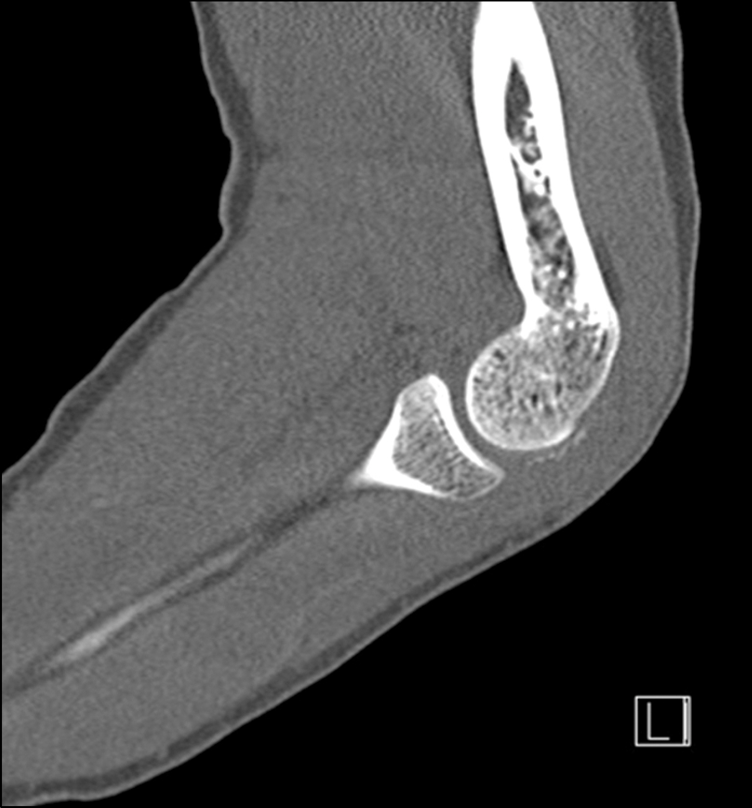
Fig. 2. In computed tomography view, small bony fragment is detected posterior to the right humerus capitulum.

Fig. 3. (A, B) In right elbow magnetic resonance imaging sagittal view, there is high signal change in ant. articular capsule and brachialis all layers. Also in axial view, it shows hematoma in brachilalis muscle and emptying sign on ligament attachment site.
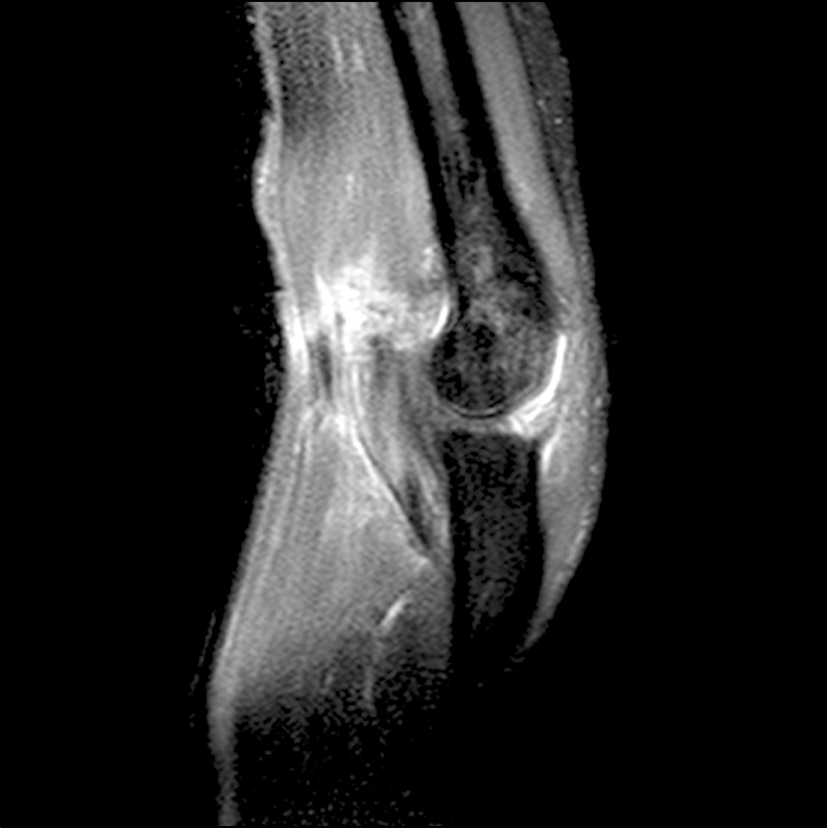
Fig. 4. In the magnetic resonance imaging sagittal view which took after a month from the event, it shows reduced hematoma and decreased signal changes around brachilalis muscle compare to previous film.

Fig. 5. In the magnetic resonance imaging sagittal view which took after 3 month from the event it shows removed hematoma and more decreased signal changes around brachilalis muscle compare to previous film.
Reference
-
1. Mehlhoff TL, Noble PC, Bennett JB, Tullos HS. Simple dislocation of the elbow in the adult. Results after closed treatment. J Bone Joint Surg Am. 1988; 70:244–9.
Article2. Mezera K, Hotchkiss RN. Fractures and dislocations of the elbow. Bucholz RW, Heckman JD, editors. editors.Rockwood and Green's fractures in adults. 5th ed.Philadelphia: Lippincott, Williams & Wilkins;2001. p. 921–34.3. Van den Berghe GR, Queenan JF, Murphy DA. Isolated rupture of the brachialis: a case report. J Bone Joint Surg Am. 2001; 83:1074–5.4. Nishida Y, Tsukushi S, Yamada Y, Hosono K, Ishiguro N. Brachialis muscle tear mimicking an intramuscular tumor: a report of two cases. J Hand Surg Am. 2007; 32:1237–41.
Article5. Winblad JB, Escobedo E, Hunter JC. Brachialis muscle rupture and hematoma. Radiol Case Rep. 2008; 3:251.
Article6. Wasserstein D, White L, Theodoropoulos J. Traumatic brachialis muscle injury by elbow hyperextension in a professional hockey player. Clin J Sport Med. 2010; 20:211–2.
Article7. Krych AJ, Kohen RB, Rodeo SA, Barnes RP, Warren RF, Hotchkiss RN. Acute brachialis muscle rupture caused by closed elbow dislocation in a professional American football player. J Shoulder Elbow Surg. 2012; 21:e1–5.
Article8. Chen HW, Chew FS. Complete rupture of both heads of the biceps brachii muscle belly by blunt trauma. Radiol Case Rep. 2006; 1:28.
Article
- Full Text Links
-
- Actions
-
Cited
- CITED
-
- Close
- Share
-
- Similar articles
-
- Chronic Median Nerve Entrapment After Posterior Fracture-Dislocation of the Elbow in a Chlid: A Case Report
- Reconstruction of the coronoid process using a graft from the olecranon of the same side for chronic posterior dislocation of the elbow
- Brachial Artery Transection with Closed Elbow Dislocation: A Case Report
- Recurrent Posterior Dislocation of the Elbow
- Posterior Dislocation of Elbow Joint with Fracture of Radial Head and Coronoid Process (Terrible triad of Elbow Fracture and Dislocation): case report
