Korean J Sports Med.
2012 Jun;30(1):9-15. 10.5763/kjsm.2012.30.1.9.
Immediate Clinical and Biomechanical Effects of Low-dye Taping in Patients with Plantar Heel Pain
- Affiliations
-
- 1Department of Physical and Rehabilitation Medicine, Samsung Medical Center, Sungkyunkwan University School of Medicine, Seoul, Korea. jhlee.hwang@samsung.com
- 2Department of Rehabilitation Medicine, Pusan National University Yangsan Hospital, Yangsan, Korea.
- KMID: 2288643
- DOI: http://doi.org/10.5763/kjsm.2012.30.1.9
Abstract
- Plantar heel pain is common musculoskeletal disorder of the foot related to sports activity. Treatment of the plantar heel pain is usually conservative including low-dye (LD) taping. We evaluated the immediate clinical and biomechanical effect of LD taping. 19 patients who had plantar heel pain with fat pad tenderness or tenderness on plantar fascia insertion area participated in this study. We assessed plantar pressure change with foot pressure analysis system, fat pad depth changes with ultrasonography, pain improvement with visual analogue scale before and after LD taping. Patient treated with LD taping showed the decrease in maximum peak pressure and pressure time integral, and there was not a significant difference between pre and post maximal velocity, average velocity, distance of center of pressure. Fat pad depth increase (mean 1.67 mm, p<0.05) and pain improvement (mean 1.91 on visual analog scale, p<0.05). LD taping restrict midtarsal joint, correct hindfoot pronation, and provide fat pad depth increase and pain improvement, immediately.
Figure
-
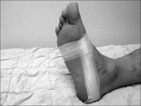
Fig. 1 Low dye taping technique.
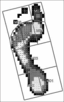
Fig. 2 The foot is divided into 8 areas. M1: medial rearfoot, M2: lateral rearfoot, M3: midfoot, M4: medial forefoot, M5: middle forefoot, M6: lateral forefoot, M7: 1st toe, M8: 2nd-5th toe.
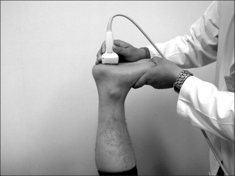
Fig. 3 The picture of weight application in ultrasonographic measurement of heel pad thickness.
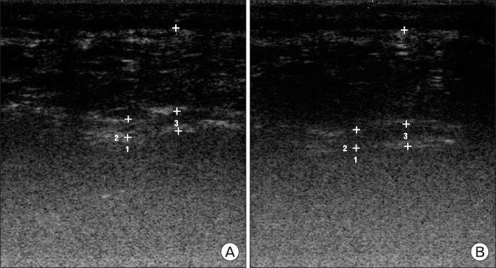
Fig. 4 (A) Thickness of heel pad in un-taped. (B) Thickness of heel pad in taped.
Cited by 1 articles
-
Clinical and Biomechanical Effects of Low-Dye Taping and Figure-8 Modification of Low-Dye Taping in Patients With Heel Pad Atrophy
You Hyeon Chae, Joo Sup Kim, Yeon Kang, Hyun Young Kim, Tae Im Yi
Ann Rehabil Med. 2018;42(2):222-228. doi: 10.5535/arm.2018.42.2.222.
Reference
-
1. Toomey EP. Plantar heel pain. Foot Ankle Clin. 2009. 14:229–245.2. Turner W, Merriman L. Clinical skills in treating the foot. 2005. 2nd ed. New York: Elsevier Churchill Livingstone.3. Chang CC, Milter LJ. Periostitis of the os calcis. J Bone Joint Surg Am. 1934. 16:355–364.4. Riddle DL, Pulisic M, Pidcoe P, Johnson RE. Risk factors for Plantar fasciitis: a matched case-control study. J Bone Joint Surg Am. 2003. 85:872–877.5. Porter D, Barrill E, Oneacre K, May BD. The effects of duration and frequency of Achilles tendon stretching on dorsiflexion and outcome in painful heel syndrome: a randomized, blinded, control study. Foot Ankle Int. 2002. 23:619–624.6. Donley BG, Moore T, Sferra J, Gozdanovic J, Smith R. The efficacy of oral nonsteroidal anti-inflammatory medication (NSAID) in the treatment of plantar fasciitis: a randomized, prospective, placebo-controlled study. Foot Ankle Int. 2007. 28:20–23.7. Miller RA, Torres J, McGuire M. Efficacy of first-time steroid injection for painful heel syndrome. Foot Ankle Int. 1995. 16:610–612.8. Pfeffer G, Bacchetti P, Deland J, et al. Comparison of custom and prefabricated orthoses in the initial treatment of proximal plantar fasciitis. Foot Ankle Int. 1999. 20:214–221.9. Lange B, Chipchase L, Evans A. The effect of low-Dye taping on plantar pressures, during gait, in subjects with navicular drop exceeding 10 mm. J Orthop Sports Phys Ther. 2004. 34:201–209.10. Nolan D, Kennedy N. Effects of low-dye taping on plantar pressure pre and post exercise: an exploratory study. BMC Musculoskelet Disord. 2009. 10:40.11. O'Sullivan K, Kennedy N, O'Neill E, Ni Mhainin U. The effect of low-dye taping on rearfoot motion and plantar pressure during the stance phase of gait. BMC Musculoskelet Disord. 2008. 9:111.12. Russo SJ, Chipchase LS. The effect of low-Dye taping on peak plantar pressures of normal feet during gait. Aust J Physiother. 2001. 47:239–244.13. Uzel M, Cetinus E, Bilgic E, Ekerbicer H, Karaoguz A. Comparison of ultrasonography and radiography in assessment of the heel pad compressibility index of patients with plantar heel pain syndrome. Measurement of the fat pad in plantar heel pain syndrome. Joint Bone Spine. 2006. 73:196–199.14. Radford JA, Landorf KB, Buchbinder R, Cook C. Effectiveness of low-Dye taping for the short-term treatment of plantar heel pain: a randomised trial. BMC Musculoskelet Disord. 2006. 7:64.15. Alexander IJ. The foot examination and diagnosis. 1997. 2nd ed. New York: Churchill Livingstone.16. Hwang JH, Chung SH. Conservative management of plantar heel pain. J Korean Acad Rehabil Med. 1998. 22:692–697.17. Hintermann B, Nigg BM. Pronation in runners. Implications for injuries. Sports Med. 1998. 26:169–176.18. Karr SD. Subcalcaneal heel pain. Orthop Clin North Am. 1994. 25:161–175.19. Gooding GA, Stress RM, Graf PM, Grunfeld C. Heel pad thickness: determination by high-resolution ultrasonography. J Ultrasound Med. 1985. 4:173–174.20. Prichasuk S. The heel pad in plantar heel pain. J Bone Joint Surg Br. 1994. 76:140–142.21. Rome K, Campbell R, Flint A, Haslock I. Reliability of weight-bearing heel pad thickness measurements by ultrasound. Clin Biomech (Bristol, Avon). 1998. 13:374–375.22. Kelly AM. Does the clinically significant difference in visual analog scale pain scores vary with gender, age, or cause of pain? Acad Emerg Med. 1998. 5:1086–1090.
- Full Text Links
-
- Actions
-
Cited
- CITED
-
- Close
- Share
-
- Similar articles
-
- Clinical and Biomechanical Effects of Low-Dye Taping and Figure-8 Modification of Low-Dye Taping in Patients With Heel Pad Atrophy
- Effect of Weight-bearing Pattern and Calcaneal Taping on Heel Width and Plantar Pressure in Standing
- Clinical Characteristics of the Causes of Plantar Heel Pain
- A Subcalcaneal Bursitis Developed after Execessive Walking Exercise
- The Effects of Modified Low-Dye Taping in the Patient with Heel Pad Atrophy
