Cluster-like Headache Secondary to Cerebral Venous Thrombosis
- Affiliations
-
- 1Department of Neurology, Seoul National University, College of Medicine, Clinical Research Institute, Seoul National University Hospital, Seoul, Korea. kimmanho@snu.ac.kr
- 2Department of Neurology, Eulji University School of Medicine, Seoul, Korea.
- KMID: 2287706
- DOI: http://doi.org/10.3988/jcn.2006.2.1.70
Abstract
- Cluster headache (CH) is considered a primary headache syndrome. However, symptomatic cases that resemble CH have also been reported. A patient with cerebral venous thrombosis presented with ipsilateral frontal pain accompanied by ophthalmoparesis, nasal congestion, and lacrimation. The patient's headache showed a dramatic response to oxygen. He experienced no further cluster-like headaches after treatment with an anticoagulant. This case suggests the possible role of venous stasis of the cavernous sinus in cluster-like headache.
MeSH Terms
Figure
-
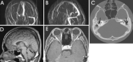
Figure 1 Images during the first admission. Brain MR venography revealed (a) no delineation of the transverse sinus on the right side and (b) tapered narrowing of the straight sinus. (c) T1-weighted image demonstrates slightly increased signal intensity (SI) or no signal void in the straight sinus. (d) Fat-suppression T1-weighted image shows no evidence of enhanced abnormal lesions in the cavernous sinus and intra-orbital structures. (e) Arrows indicate mastoid air cells with soft tissue opacity on both sides and air-fluid levels on the left side, which are consistent with mastoiditis of both sides.

Figure 2 Images during the second admission with seizure. (A) T2-weighted image shows hyperintense lesions in the left parietotemporal area. (B) A heterogeneous SI lesion is evident on the diffusion-weighted image. An isointensity signal of the clot in the superior sagittal sinus is also evident. (C) An apparent diffusion coefficient map reveals mixed intensities with high and low SIs at comparable sites. (D, E) Contrast-enhanced MR venography shows a poorly visualized superior sagittal sinus that suggests thrombosis as well as persistent venous thrombosis involving both the straight and right transverse sinuses.
Reference
-
1. Moskcowitz MA. Cluster headache: evidence for a pathophysiologic focus in the superior pericarotid cavernous sinus plexus. Headache. 1988. 28:584–586.
Article2. Hannerz J, Ericson K, Bergstrand G. Orbital phlebography in patients with cluster headache. Cephalalgia. 1987. 7:207–211.
Article3. Hannerz J. A case of parasellar meningioma mimicking cluster headache. Cephalalgia. 1989. 9:265–269.
Article4. Todo T, Inoya H. Sudden appearance of a mycotic aneurysm of the intracavernous carotid artery after symptoms resembling cluster headache: case report. Neurosurgery. 1991. 29:594–599.
Article5. May A, Bahra A, Buchel C, Frackowiak RSJ, Goadsby PJ. Hypothalamic activation in cluster attacks. Lancet. 1998. 352:275–278.6. Leone M, Bussone G. A review of hormonal findings in cluster headache. Evidence for hypothalamic involvement. Cephalalgia. 1993. 13:309–317.
Article7. Frishberg BM. Neuroimaging in presumed primary headache disorders. Semin Neurol. 1997. 17:373–382.
Article8. Ameri A, Bousser MG. Cerebral venous thrombosis. Neurol Clin. 1992. 10:87–111.
Article9. Afra J, Cecchini AP, Schoenen J. Craniometric measures in cluster headache patients. Cephalalgia. 1998. 18:143–145.
Article10. Hannerz J, Jogestrand T. Chronic cluster headache: provocation with carbon dioxide breathing and nitroglycerin. Headache. 1996. 36:174–177.
Article
- Full Text Links
-
- Actions
-
Cited
- CITED
-
- Close
- Share
-
- Similar articles
-
- Parasellar Meningioma Mimicking Cluster Headache Treated with Novalis Stereotactic Radiosurgery
- Cerebral Venous Thrombosis Complicated by Intracranial Hypotension
- Deep Cerebral Venous Thrombosis Showing Parkinsonism such as Micrographia, Hypophonia and Bradykinesia
- A Case of Deep Cerebral Venous Thrombosis Associated with Iron Deficiency Anemia
- A Case of Leptomeningeal Metastasis Associated with Cerebral Venous Thrombosis
