Classification and Serial Evolution of PLEDs
- Affiliations
-
- 1Department of Neurology, Chungnam National University College of Medicine, Daejeon, Korea. jmoonkim@cnu.ac.kr
- KMID: 2287691
- DOI: http://doi.org/10.3988/jcn.2006.2.3.179
Abstract
- BACKGROUND AND PURPOSE
Periodic lateralized epileptiform discharges (PLEDs) are defined as spikes or sharp waves occurring at an approximately regular interval. PLEDs are subdivided into PLEDs proper and PLEDs plus in Reiher's classification, but since this does not sufficiently reflect the pleomorphism of PLEDs, we propose a new subclassification scheme of PLEDs, and discuss the relationship between them and clinical prognoses.
METHODS
Thirty-seven patients who had at least two available EEGs were included in this study. Each patient had structural brain lesions identified in brain CT/MRI: 237 EEGs from 37 patients were reviewed and the patterns of PLEDs were classified by electroencephalographic characteristics based on Reiher's classification. PLEDs proper of class 3 were subclassified into four categories: (1) simple, (2) benign, (3) vigorous, and (4) suppressed.
RESULTS
Most of the PLEDs that started with the vigorous or suppressed pattern of class 3 evolved into the simple or benign pattern of class 3 and subsequently changed into class 1 or class 2, finally intermingling with the neighboring background waves. PLEDs that started with the benign or simple pattern of class 3 rapidly changed into class 1 or 2. Patients showing the benign or simple pattern of class 3 exhibited a better clinical prognosis.
CONCLUSIONS
PLEDs have five distinctive classes, and over time they evolve from malignant PLEDs plus to benign PLEDs proper before finally disappearing. It appears that those of class 3 have more diverse patterns, with the vigorous and suppressed patterns being the more malignant forms of PLEDs in this class.
Keyword
Figure
-

Figure 1 Representative EEG recordings showing diverse patterns of PLEDs proper of class 3: (A) simple, (B) benign, (C) vigorous, and (D) suppressed.

Figure 2 Average evolution of five patients with initial vigorous PLEDs proper of class 3. The X-axis represents the sequence of EEG recordings and the Y-axis represents the EEG findings: 1; slowing, 2; occasional sharp, 3; frequent sharp, 4; PLEDs proper of class 1, 5; PLEDs proper of class 2, 6; simple PLEDs proper of class 3, 7; benign PLEDs proper of class 3, 8; vigorous PLEDs proper of class 3, 9; suppressed PLEDs proper of class 3, 10; PLEDs plus of class 4, and 11; PLEDs plus of class 5.
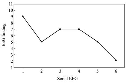
Figure 3 Average evolution of two patients with initial suppressed PLEDs proper of class 3 (axes as in Fig. 2).
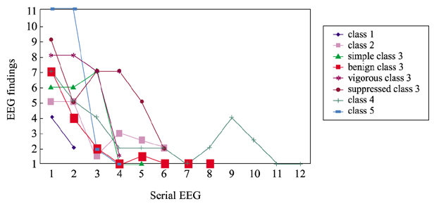
Figure 4 Stepwise evolution of PLEDs (axes as in Fig. 2).

Figure 5 Relationship between PLEDs classification and functional status at follow-up: (A) benign group comprising PLEDs proper of classes 1 and 2, and simple or benign PLEDs proper of class 3 (n=25), and (B) malignant group comprising vigorous or suppressed PLEDs proper of class 3, and PLEDs plus of class or 5 (n=17).
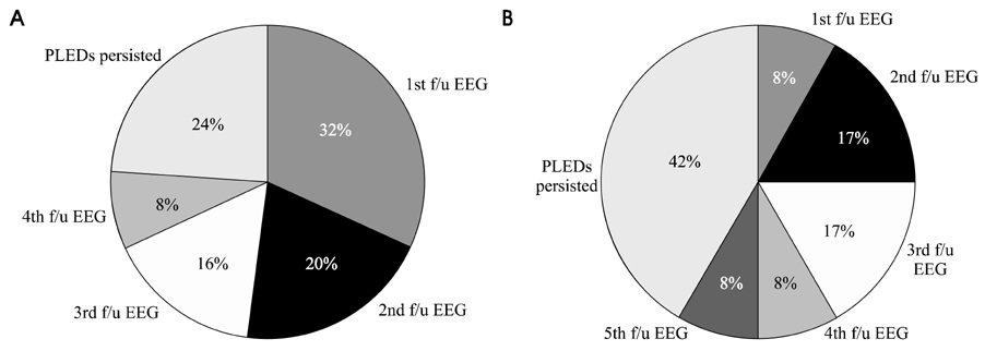
Figure 6 Resolution of PLEDs during the follow-up EEGs: (A) benign group and (B) malignant group. f/u; follow-up.

Figure 7 EEG seizure and functional status at follow-up: (A) with EEG seizure (n=7) and (B) without EEG seizure (n=30).
Reference
-
1. Chatrian GE, Shaw CM, Leffman H. The significance of periodic lateralized epileptiform discharges in EEG: an electrographic, clinical and pathological study. Electroencephalogr Clin Neurophysiol. 1964. 17:177–193.
Article2. Kuroiwa Y, Celesia GG. Clinical significance of periodic EEG patterns. Arch Neurol. 1980. 37:15–20.
Article3. Brenner RP, Schaul N. Periodic EEG patterns: classification, clinical correlation, and pathophysiology. J Clin Neurophysiol. 1990. 7:249–267.4. Hirsch LJ, Claassen J, Mayer SA, Emerson RG. Stimulusinduced rhythmic, periodic, or ictal discharges (SIRPIDs): a common EEG phenomenon in the critically ill. Epilepsia. 2004. 45:109–123.
Article5. Reiher J, Rivest J, Grand' Maison F, Leduc CP. Periodic lateralized epileptiform discharges with transitional rhythmic discharges: association with seizures. Electroencephalogr Clin Neurophysiol. 1991. 78:12–17.
Article6. Garcia-Morales I, Garcia MT, Galan-Davila L, Gomez-Escalonilla C, Saiz-Diaz R, Martinez-Salio A, et al. Periodic lateralized epileptiform discharges: etiology, clinical aspects, seizures, and evolution in 130 patients. J Clin Neurophysiol. 2002. 19:172–177.7. Pohlmann-Eden B, Hoch DB, Cochius JI, Chiappa KH. Periodic lateralized epileptiform discharges - a critical review. J Clin Neurophysiol. 1996. 13:519–530.8. Walsh JM, Brenner RP. Periodic lateralized epileptiform discharges - long-term outcome in adults. Epilepsia. 1987. 28:533–536.
Article9. Treiman DM, Walton NY, Kendrick C. A progressive sequence of electroencephalographic changes during generalized convulsive status epilepticus. Epilepsy Res. 1990. 5:49–60.
Article10. Baykan B, Kinay D, Gokyigit A, Gurses C. Periodic lateralized epileptiform discharges: association with seizures. Seizure. 2000. 9:402–406.
Article11. Snodgrass SM, Tsuburaya K, Ajmone-Marsan C. Clinical significance of periodic lateralized epileptiform discharges: relationship with status epilepticus. J Clin Neurophysiol. 1989. 6:159–172.12. Chong DJ, Hirsch LJ. Which EEG patterns warrant treatment in the critically ill? Reviewing the evidence for treatment of periodic lateralized epileptiform discharges and related patterns. J Clin Neurophysiol. 2005. 22:79–91.
Article13. Silverman IE, Restrepo L, Mathews GC. Poststroke seizures. Arch Neurol. 2002. 59:195–201.
Article
- Full Text Links
-
- Actions
-
Cited
- CITED
-
- Close
- Share
-
- Similar articles
-
- Periodic lateralized epileptiform discharges(PLEDs) with partial seizures after alcohol withdrawal
- A Study on the Clinical Significance of Periodic Lateralized Epileptiform Discharges and Relation to Brain Imaging Study in Children
- Current Source Distribution of Periodic Lateralized Epileptiform Discharge: Comparison With Diffusion-Wighted MR Imaging in Viral Encephalitis
- Molecular Epidemiology of Human Immunodeficiency Virus
- Quantitative EEG Characteristics of Periodic Lateralized Epileptiform Discharges according to Benzodiazepine Responsiveness
