J Clin Neurol.
2012 Mar;8(1):43-50. 10.3988/jcn.2012.8.1.43.
Medial Temporal Atrophy and Memory Dysfunction in Poststroke Cognitive Impairment-No Dementia
- Affiliations
-
- 1Department of Neurology, Seoul National University Hospital, Seoul National University College of Medicine, Seoul, Korea.
- 2Department of Neurology, Seoul National University Bundang Hospital, Seoul National University College of Medicine, Seongnam, Korea. braindoc@snu.ac.kr
- 3Department of Biostatistics, Korea University College of Medicine, Seoul, Korea.
- 4Department of Psychology, Hallym University, Chuncheon, Korea.
- 5Department of Neurology, Hallym University College of Medicine, Chuncheon, Korea.
- KMID: 2287616
- DOI: http://doi.org/10.3988/jcn.2012.8.1.43
Abstract
- BACKGROUND AND PURPOSE
It was recently reported that the prevalence of poststroke memory dysfunction might be higher than previously thought. Stroke may exist concomitantly with underlying Alzheimer's disease (AD), and so we determined whether post-stroke memory dysfunction indicates manifestation of underlying subclinical AD.
METHODS
Of 1201 patients in a prospective cognitive assessment database, we enrolled subjects with poststroke amnestic vascular cognitive impairment-no dementia (aVCIND; n=48), poststroke nonamnestic vascular cognitive impairment-no dementia (naVCIND; n=50), and nonstroke amnestic mild cognitive impairment (aMCI; n=65). All subjects had cognitive deficits, but did not meet the criteria for dementia. A standardized neuropsychological test battery and magnetic resonance imaging were performed at least 90 days after the index stroke (mean, 473 days). Visual assessment of medial temporal atrophy (MTA) was used as a measure of underlying AD pathology.
RESULTS
The MTA score was significantly lower in the naVCIND group (0.64+/-0.85, mean+/-SD) than in the aVCIND (1.10+/-1.08) and aMCI (1.45+/-1.13; p<0.01) groups. Multivariable ordinal logistic regression analysis revealed that compared with naVCIND, aVCIND [odds ratio (OR)=2.69; 95% confidence interval (CI)=1.21-5.99] and aMCI (OR=5.20; 95% CI=2.41-11.23) were significantly associated with increasing severity of MTA.
CONCLUSIONS
Our findings show that compared with poststroke naVCIND, the odds of having more-severe MTA were increased for poststroke aVCIND and nonstroke aMCI.
MeSH Terms
Figure
-
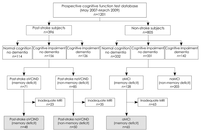
Fig. 1 Recruitment of the study population. Gray boxes denote the enrolled subjects who were included in the final analyses. aVCIND: amnestic vascular cognitive impairment-no dementia, na-VCIND: nonamnestic vascular cognitive impairment-no dementia, aMCI: amnestic mild cognitive impairment.
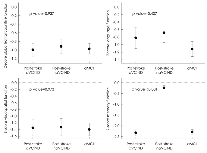
Fig. 2 Comparison of Z-scores among the three vascular cognitive impairment and no dementia (CIND) groups. aMCI: amnestic mild cognitive impairment, aVCIND: amnestic vascular cognitive impairment-no dementia, naVCIND: nonamnestic vascular cognitive impairment-no dementia.
Reference
-
1. Hachinski V. The 2005 Thomas Willis Lecture: stroke and vascular cognitive impairment: a transdisciplinary, translational and transactional approach. Stroke. 2007. 38:1396.2. Kavirajan H, Schneider LS. Efficacy and adverse effects of cholinesterase inhibitors and memantine in vascular dementia: a meta-analysis of randomised controlled trials. Lancet Neurol. 2007. 6:782–792.
Article3. Traykov L, Baudic S, Thibaudet MC, Rigaud AS, Smagghe A, Boller F. Neuropsychological deficit in early subcortical vascular dementia: comparison to Alzheimer's disease. Dement Geriatr Cogn Disord. 2002. 14:26–32.
Article4. Snaphaan L, de Leeuw FE. Poststroke memory function in nondemented patients: a systematic review on frequency and neuroimaging correlates. Stroke. 2007. 38:198–203.5. Kalaria RN. The role of cerebral ischemia in Alzheimer's disease. Neurobiol Aging. 2000. 21:321–330.
Article6. Rockwood K, Wentzel C, Hachinski V, Hogan DB, MacKnight C, McDowell I. Prevalence and outcomes of vascular cognitive impairment. Vascular Cognitive Impairment Investigators of the Canadian Study of Health and Aging. Neurology. 2000. 54:447–451.
Article7. Korf ES, Wahlund LO, Visser PJ, Scheltens P. Medial temporal lobe atrophy on MRI predicts dementia in patients with mild cognitive impairment. Neurology. 2004. 63:94–100.
Article8. WHO MONICA Project Principal Investigators. The World Health Organization MONICA Project (monitoring trends and determinants in cardiovascular disease): a major international collaboration. J Clin Epidemiol. 1988. 41:105–114.9. American Psychiatric Association. Diagnostic and Statistical Manual of Mental Disorders: DSM-IV-TR. 2000. 4th ed, text revision. Washington, DC: American Psychiatric Association.10. Kang SJ, Choi SH, Lee BH, Kwon JC, Na DL, Han SH. Korean Dementia Research Group. The reliability and validity of the Korean instrumental activities of daily living (K-IADL). J Korean Neurol Assoc. 2002. 20:8–14.11. Hachinski V, Iadecola C, Petersen RC, Breteler MM, Nyenhuis DL, Black SE, et al. National Institute of Neurological Disorders and Stroke-Canadian Stroke Network vascular cognitive impairment harmonization standards. Stroke. 2006. 37:2220–2241.
Article12. Kang Y. Recent developments in the Korean VCI neuropsychology harmonization efforts. 2009. In : The 4th Congress of the International Society for Vascular Behavioural and Cognitive Disorders (VAS-COG); Singapore.13. Scheltens P, Leys D, Barkhof F, Huglo D, Weinstein HC, Vermersch P, et al. Atrophy of medial temporal lobes on MRI in "probable" Alzheimer's disease and normal ageing: diagnostic value and neuropsychological correlates. J Neurol Neurosurg Psychiatry. 1992. 55:967–972.
Article14. Scheltens P, Launer LJ, Barkhof F, Weinstein HC, van Gool WA. Visual assessment of medial temporal lobe atrophy on magnetic resonance imaging: interobserver reliability. J Neurol. 1995. 242:557–560.
Article15. Wong TY, Klein R, Sharrett AR, Couper DJ, Klein BE, Liao DP, et al. Cerebral white matter lesions, retinopathy, and incident clinical stroke. JAMA. 2002. 288:67–74.
Article16. Wagner AD, Schacter DL, Rotte M, Koutstaal W, Maril A, Dale AM, et al. Building memories: remembering and forgetting of verbal experiences as predicted by brain activity. Science. 1998. 281:1188–1191.
Article17. Savva GM, Stephan BC. Alzheimer's Society Vascular Dementia Systematic Review Group. Epidemiological studies of the effect of stroke on incident dementia: a systematic review. Stroke. 2010. 41:e41–e46.18. Staekenborg SS, Koedam EL, Henneman WJ, Stokman P, Barkhof F, Scheltens P, et al. Progression of mild cognitive impairment to dementia: contribution of cerebrovascular disease compared with medial temporal lobe atrophy. Stroke. 2009. 40:1269–1274.
Article19. Niwa K, Kazama K, Younkin L, Younkin SG, Carlson GA, Iadecola C. Cerebrovascular autoregulation is profoundly impaired in mice over-expressing amyloid precursor protein. Am J Physiol Heart Circ Physiol. 2002. 283:H315–H323.20. Iadecola C. Neurovascular regulation in the normal brain and in Alzheimer's disease. Nat Rev Neurosci. 2004. 5:347–360.
Article21. Selden NR, Gitelman DR, Salamon-Murayama N, Parrish TB, Mesulam MM. Trajectories of cholinergic pathways within the cerebral hemispheres of the human brain. Brain. 1998. 121:2249–2257.
Article22. Román GC, Kalaria RN. Vascular determinants of cholinergic deficits in Alzheimer disease and vascular dementia. Neurobiol Aging. 2006. 27:1769–1785.
Article23. Fratiglioni L, Paillard-Borg S, Winblad B. An active and socially integrated lifestyle in late life might protect against dementia. Lancet Neurol. 2004. 3:343–353.
Article24. Viswanathan A, Rocca WA, Tzourio C. Vascular risk factors and dementia: how to move forward? Neurology. 2009. 72:368–374.
Article25. Zekry D. Is it possible to treat vascular dementia? Front Neurol Neurosci. 2009. 24:95–106.
Article26. Hénon H, Pasquier F, Durieu I, Pruvo JP, Leys D. Medial temporal lobe atrophy in stroke patients: relation to pre-existing dementia. J Neurol Neurosurg Psychiatry. 1998. 65:641–647.
Article27. Pohjasvaara T, Mäntylä R, Salonen O, Aronen HJ, Ylikoski R, Hietanen M, et al. MRI correlates of dementia after first clinical ischemic stroke. J Neurol Sci. 2000. 181:111–117.
Article28. Jokinen H, Kalska H, Ylikoski R, Hietanen M, Mäntylä R, Pohjasvaara T, et al. Medial temporal lobe atrophy and memory deficits in elderly stroke patients. Eur J Neurol. 2004. 11:825–832.
Article29. Van der Werf YD, Scheltens P, Lindeboom J, Witter MP, Uylings HB, Jolles J. Deficits of memory, executive functioning and attention following infarction in the thalamus; a study of 22 cases with localised lesions. Neuropsychologia. 2003. 41:1330–1344.
Article30. den Heijer T, Vermeer SE, van Dijk EJ, Prins ND, Koudstaal PJ, Hofman A, et al. Type 2 diabetes and atrophy of medial temporal lobe structures on brain MRI. Diabetologia. 2003. 46:1604–1610.
Article31. van de Pol LA, Verhey F, Frisoni GB, Tsolaki M, Papapostolou P, Nobili F, et al. White matter hyperintensities and medial temporal lobe atrophy in clinical subtypes of mild cognitive impairment: the DESCRIPA study. J Neurol Neurosurg Psychiatry. 2009. 80:1069–1074.
Article32. Appel J, Potter E, Bhatia N, Shen Q, Zhao W, Greig MT, et al. Association of white matter hyperintensity measurements on brain MR imaging with cognitive status, medial temporal atrophy, and cardiovascular risk factors. AJNR Am J Neuroradiol. 2009. 30:1870–1876.
Article33. Moorhouse P, Song X, Rockwood K, Black S, Kertesz A, Gauthier S, et al. Executive dysfunction in vascular cognitive impairment in the consortium to investigate vascular impairment of cognition study. J Neurol Sci. 2010. 288:142–146.
Article34. Kelley BJ, Petersen RC. Miller BL, Boeve BF, editors. Mild cognitive impairment. The behavioral neurology of dementia. 2009. New York: Cambridge University Press;172–187.
Article35. Wahlund LO, Julin P, Johansson SE, Scheltens P. Visual rating and volumetry of the medial temporal lobe on magnetic resonance imaging in dementia: a comparative study. J Neurol Neurosurg Psychiatry. 2000. 69:630–635.
Article36. DeCarli C, Frisoni GB, Clark CM, Harvey D, Grundman M, Petersen RC, et al. Qualitative estimates of medial temporal atrophy as a predictor of progression from mild cognitive impairment to dementia. Arch Neurol. 2007. 64:108–115.
Article37. Erkinjuntti T, Inzitari D, Pantoni L, Wallin A, Scheltens P, Rockwood K, et al. Limitations of clinical criteria for the diagnosis of vascular dementia in clinical trials. Is a focus on subcortical vascular dementia a solution? Ann N Y Acad Sci. 2000. 903:262–272.
Article38. Hong KS, Kim J, Cho YJ, Seo SY, Hwang SI, Kim SC, et al. Burden of ischemic stroke in Korea: analysis of disability-adjusted life years lost. J Clin Neurol. 2011. 7:77–84.
Article
- Full Text Links
-
- Actions
-
Cited
- CITED
-
- Close
- Share
-
- Similar articles
-
- Dementia due to Meningovascular Syphilis in Medial Temporal Lobe and Cognitive Rehabilitation
- Can We Further Divide Amnestic Mild Cognitive Impairment Based on the Pattern of Memory Deficit?: A Preliminary Study
- Memory Impairment in Dementing Patients
- Medial Temporal Atrophy Alone is Insufficient to Predict Underlying Alzheimer’s Disease Pathology
- Epidemiology of Age-Associated Memory Impairment
