Acute Spinal-Cord Ischemia: Evolution of MRI Findings
- Affiliations
-
- 1Department of Neurology, Medical Center Haaglanden, The Hague, The Netherlands. calblas@vlietlandziekenhuis.nl
- 2Department of Radiology, Medical Center Haaglanden, The Hague, The Netherlands.
- KMID: 2287594
- DOI: http://doi.org/10.3988/jcn.2012.8.3.218
Abstract
- BACKGROUND AND PURPOSE
Magnetic resonance (MR) findings in acute spinal-cord ischemia can be summarized as focal cord enlargement and hyperintensities on T2-weighted images and gadolinium enhancement, especially of the central gray matter. However, in analogy with acute brain ischemia, it is to be expected that the findings of MR imaging (MRI) may be normal in the first hours after symptom onset. We evaluated the clinical and MRI findings in a series of patients with acute spinal-cord ischemia, and tested the hypothesis that the development and course of MR abnormalities are predictable.
METHODS
Five patients with acute spinal-cord ischemia were admitted to our hospital over a 2-year period. Repeated MRI (1.5 T) was performed in all patients. Clinical data were retrieved from the patients' charts.
RESULTS
Four women and one man with a median age of 52 years (range, 31-75 years) were admitted. Three patients had anterior spinal artery infarction and two patients had transverse infarctions. All patients underwent spinal MRI within 24 hours; the findings were normal in four of the five patients. After 1-2 days, T2-weighted MRI generally exhibited focal cord enlargement and hyperintensity in all patients, while spinal-cord enhancement appeared after 2-11 days.
CONCLUSIONS
Acute spinal-cord ischemia may have a typical course on MRI. MRI findings are usually normal in the acute phase, but spinal cord swelling and T2 abnormality are expected after several days, while gadolinium enhancement appears even later after symptom onset. The sensitivity and specificity of MRI can be increased by repeated MRI in patients suspected of acute spinal-cord ischemia.
Keyword
MeSH Terms
Figure
-
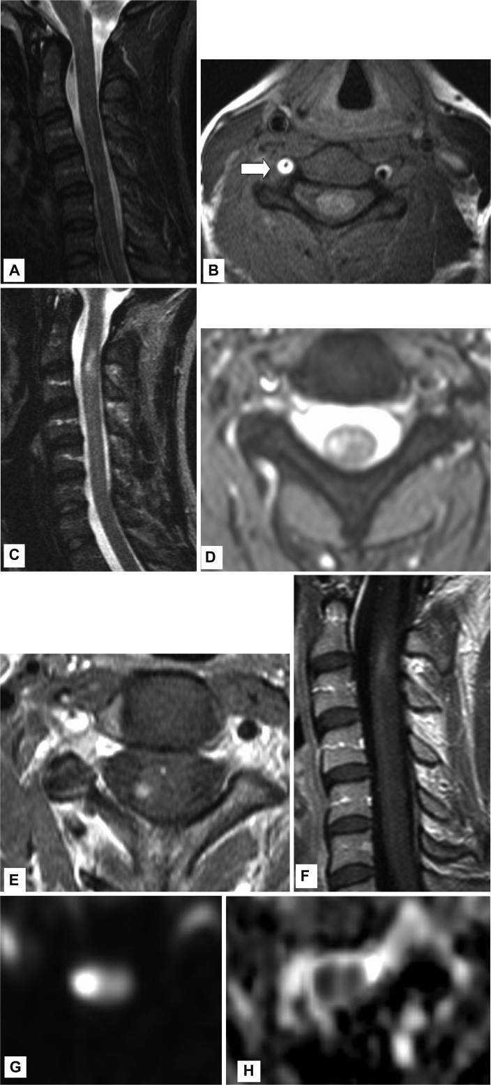
Fig. 1 A: MRI performed 12 hours after symptom onset: 31-year-old female presenting with right-sided Horner syndrome and right-sided loss of strength. Sagittal T2-weighted image showing no cord abnormalities. B: MRI performed 12 hours after symptom onset: axial T1-weighted image with fat suppression showing an abnormally high signal intensity (SI) in most of the cross-sectional area of the right vertebral artery, with a small central flow void (arrow), confirming suspected vertebral artery dissection. C and D: MRI performed 6 days after symptom onset: sagittal (C) and axial (D) T2-weighted images showing abnormal areas of high SI on the right at the C2 level.E and F: MRI performed 6 days after symptom onset: axial (E) and sagittal (F) T1-weighted images after administration of gadolinium, revealing enhancement of the cord at the C2 level. G and H: MRI performed 6 days after symptom onset: axial (G) DWI showing right-sided high signal at the C2 level and axial (H) low apparent diffusion coefficient values showing diffusion restriction, supporting the diagnosis of spinal-cord ischemia. DWI: diffusion-weighted imaging, MRI: magnetic resonance imaging.
Cited by 1 articles
-
Atypical Anterior Spinal Artery Infarction due to Left Vertebral Artery Occlusion Presenting with Bilateral Hand Weakness
Min-Ji Kim, Mi-Hee Jang, Mi-Song Choi, Suk Yun Kang, Joo Yong Kim, Ki-Han Kwon, Ik-Won Kang, Soo-Jin Cho
J Clin Neurol. 2014;10(2):171-173. doi: 10.3988/jcn.2014.10.2.171.
Reference
-
1. de Seze J, Stojkovic T, Breteau G, Lucas C, Michon-Pasturel U, Gauvrit JY, et al. Acute myelopathies: Clinical, laboratory and outcome profiles in 79 cases. Brain. 2001; 124:1509–1521. PMID: 11459743.
Article2. Novy J, Carruzzo A, Maeder P, Bogousslavsky J. Spinal cord ischemia: clinical and imaging patterns, pathogenesis, and outcomes in 27 patients. Arch Neurol. 2006; 63:1113–1120. PMID: 16908737.3. Masson C, Pruvo JP, Meder JF, Cordonnier C, Touzé E, De La Sayette V, et al. Spinal cord infarction: clinical and magnetic resonance imaging findings and short term outcome. J Neurol Neurosurg Psychiatry. 2004; 75:1431–1435. PMID: 15377691.
Article4. Nedeltchev K, Loher TJ, Stepper F, Arnold M, Schroth G, Mattle HP, et al. Long-term outcome of acute spinal cord ischemia syndrome. Stroke. 2004; 35:560–565. PMID: 14726546.
Article5. Weidauer S, Nichtweiss M, Lanfermann H, Zanella FE. Spinal cord infarction: MR imaging and clinical features in 16 cases. Neuroradiology. 2002; 44:851–857. PMID: 12389137.
Article6. Thurnher MM, Bammer R. Diffusion-weighted MR imaging (DWI) in spinal cord ischemia. Neuroradiology. 2006; 48:795–801. PMID: 16977443.
Article7. Grossman RI, Yousem DM. Neuroradiology: The Requisites. 2003. 2nd ed. Philadelphia: Mosby;p. 831–832.8. Ross JS, Brant-Zawadzki M, Moore KR, Crim J, Chen MZ, Katzman GL. Diagnostic Imaging. Spine. 2004. Salt Lake City: Amirsys;p. 26–29.9. Sibon I, Ménégon P, Moonen CT, Dousset V. Early diagnosis of spinal cord infarct using magnetic resonance diffusion imaging. Neurology. 2003; 61:1622. PMID: 14663058.
Article10. Fujikawa A, Tsuchiya K, Takeuchi S, Hachiya J. Diffusion-weighted MR imaging in acute spinal cord ischemia. Eur Radiol. 2004; 14:2076–2078. PMID: 15022011.
Article11. Nowak DA, Mutzenbach S, Fuchs HH. Acute myelopathy. Retrospective clinical, laboratory, MRI and outcome analysis of 49 cases. J Clin Neurosci. 2004; 11:145–152. PMID: 14732373.
Article12. Thurnher MM, Bammer R. Diffusion-weighted magnetic resonance imaging of the spine and spinal cord. Semin Roentgenol. 2006; 41:294–311. PMID: 17010692.
Article13. Küker W, Weller M, Klose U, Krapf H, Dichgans J, Nägele T. Diffusion-weighted MRI of spinal cord infarction--high resolution imaging and time course of diffusion abnormality. J Neurol. 2004; 251:818–824. PMID: 15258783.
- Full Text Links
-
- Actions
-
Cited
- CITED
-
- Close
- Share
-
- Similar articles
-
- Acute Rhabdomyolysis and Ischemia of the Spinal Cord Following the Heavy Alcohol Ingestion: A case report
- Spinal Cord Ischemia Secondary to Hypovolemic Shock
- Significance of MRI Cord Signal Patterns in Acute Spinal Trauma
- Myelopathy Caused by Surfing
- Vertebral Artery Dissection Presenting with Acute Infarction in Cervical Spinal Cord and Cerebellum
