J Clin Neurol.
2014 Apr;10(2):171-173. 10.3988/jcn.2014.10.2.171.
Atypical Anterior Spinal Artery Infarction due to Left Vertebral Artery Occlusion Presenting with Bilateral Hand Weakness
- Affiliations
-
- 1Department of Neurology, Kangnam Sacred Heart Hospital, Hallym University College of Medicine, Seoul, Korea.
- 2Department of Neurology, Hallym University Sacred Heart Hospital, Anyang, Korea.
- 3Department of Neurology, Dongtan Sacred Heart Hospital, Hallym University College of Medicine, Hwaseong, Korea. dowonc@naver.com
- 4Department of Radiology, Dongtan Sacred Heart Hospital, Hallym University College of Medicine, Hwaseong, Korea.
- KMID: 2287537
- DOI: http://doi.org/10.3988/jcn.2014.10.2.171
Abstract
- BACKGROUND
Infarct of the anterior spinal artery is the most common subtype of spinal cord infarct, and is characterized by bilateral motor deficits with spinothalamic sensory deficits. We experienced a case with atypical anterior-spinal-artery infarct that presented with bilateral hand weakness but without sensory deficits.
CASE REPORT
A 29-year-old man presented with sudden neck pain and bilateral weakness of the hands. Magnetic resonance imaging (MRI) of the brain did not reveal any lesion. His motor symptoms improved rapidly except for mild weakness in his left wrist and fingers. Magnetic resonance angiography showed proximal occlusion of the left vertebral artery; a spine MRI revealed left cervical cord infarction.
CONCLUSIONS
Bilateral or unilateral hand weakness can be the sole symptom of a cervical cord infarct.
MeSH Terms
Figure
-

Fig. 1 A: Severe stenosis and occlusion of the proximal part of the left vertebral artery in a neck CT angiogram. B: Axial T2-weighted image showing severe stenosis of the left vertebral artery due to dissection, showing a pseudo lumen with mural thrombi (arrow), and a high signal intensity in the left gray matter of spinal cord at the C4 level.
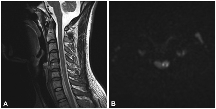
Fig. 2 A: Sagittal T2-weighted MRI image of the cervical spine showing an abnormal area of high signal intensities at the C3, C4, and C6 levels. B: Diffusion-weighted image showing high signal intensities on the left side at the C3 and C4 levels.
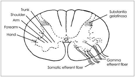
Fig. 3 Approximate locations of the motor neurons in the anterior gray horn of the cervical cord.
Reference
-
1. Novy J, Carruzzo A, Maeder P, Bogousslavsky J. Spinal cord ischemia: clinical and imaging patterns, pathogenesis, and outcomes in 27 patients. Arch Neurol. 2006; 63:1113–1120.2. Kumral E, Polat F, Güllüoglu H, Uzunköprü C, Tuncel R, Alpaydin S. Spinal ischaemic stroke: clinical and radiological findings and short-term outcome. Eur J Neurol. 2011; 18:232–239.
Article3. Satran R. Spinal cord infarction. Stroke. 1988; 19:529–532.
Article4. Berg D, Müllges W, Koltzenburg M, Bendszus M, Reiners K. Man-in-the-barrel syndrome caused by cervical spinal cord infarction. Acta Neurol Scand. 1998; 97:417–419.5. Li Y, Jenny D, Bemporad JA, Liew CJ, Castaldo J. Sulcal artery syndrome after vertebral artery dissection. J Stroke Cerebrovasc Dis. 2010; 19:333–335.
Article6. Machnowska M, Moien-Afshari F, Voll C, Wiebe S. Partial anterior cervical cord infarction following vertebral artery dissection. Can J Neurol Sci. 2008; 35:674–677.
Article7. Park SW, Sohn SI, Cho YW, Lee H, Lim JG, Yi SD. Spinal cord infarction with anterior chest pain. J Korean Neurol Assoc. 2005; 23:840–841.8. Pullicino P. Bilateral distal upper limb amyotrophy and watershed infarcts from vertebral dissection. Stroke. 1994; 25:1870–1872.
Article9. Parent A. Spinal cord: regional anatomy and internal structure. In : Parent A, editor. Carpenter's Human Neuroanatomy. 9th ed. Baltimore: Williams & Wilkins;1996. p. 325–367.10. Alblas CL, Bouvy WH, Lycklama À Nijeholt GJ, Boiten J. Acute spinal-cord ischemia: evolution of MRI findings. J Clin Neurol. 2012; 8:218–223.
Article11. Penfield W, Boldrey E. Somatic motor and sensory representation in the cerebral cortex of man studied by electrical stimulation. Brain. 1937; 60:389–443.
Article12. Dawson DM, Potts F. Acute nontraumatic myelopathies. Neurol Clin. 1991; 9:585–603.
Article13. Robertson CE, Brown RD Jr, Wijdicks EF, Rabinstein AA. Recovery after spinal cord infarcts: long-term outcome in 115 patients. Neurology. 2012; 78:114–121.
Article
- Full Text Links
-
- Actions
-
Cited
- CITED
-
- Close
- Share
-
- Similar articles
-
- Cervical Spinal Cord Infarction Presenting as Chest Pain in Patients with Acute Cerebellar Infarction
- Delayed Brain Infarction due to Bilateral Vertebral Artery Occlusion Which Occurred 5 Days after Cervical Trauma
- Vertebral Artery Dissection Presenting with Acute Infarction in Cervical Spinal Cord and Cerebellum
- A Case of Bilateral Spontaneous Extracranial Vertebral Artery Dissection
- Bilateral Thalamic Infarction Related to Artery of Percheron with Microembolic Signal
