Correlation between Ultrasonography Findings and Electrodiagnostic Severity in Carpal Tunnel Syndrome: 3D Ultrasonography
- Affiliations
-
- 1Department of Physical Medicine & Rehabilitation, Korea University Anam Hospital, Korea University College of Medicine, Seoul, Korea.
- 2Department of Physical Medicine & Rehabilitation, Korea University Guro Hospital, Korea University College of Medicine, Seoul, Korea. rehab46@korea.ac.kr
- 3Department of Radiology, Korea University Anam Hospital, Korea University College of Medicine, Seoul, Korea.
- KMID: 2287515
- DOI: http://doi.org/10.3988/jcn.2014.10.4.348
Abstract
- BACKGROUND AND PURPOSE
To determine the correlation between the cross-sectional area (CSA) of the median nerve measured at the wrist using three-dimensional (3D) ultrasonography (US) and the electrophysiological severity of carpal tunnel syndrome (CTS).
METHODS
We prospectively examined 102 wrists of 51 patients with clinical CTS, which were classified into 3 groups according to the electrodiagnostic (EDX) findings. Median nerve CSAs were measured using 3D US at the carpal tunnel inlet and at the level of maximal swelling.
RESULTS
Ten wrists were negative for CTS. Of the 92 CTS-positive wrists, 23, 30, and 39 were classified as having mild, moderate, and severe CTS, respectively. The median nerve CSA differed significantly between the severe- and moderate-CTS groups (p=0.0007 at the carpal tunnel inlet and p<0.0001 at the maximal swelling site). There was a correlation between median nerve CSA and EDX parameters among those wrists with severe and mild CTS (p<0.0001 at both sites).
CONCLUSIONS
The median nerve CSA as measured by 3D US could provide additional information about the severity of CTS, as indicated by the strong correlation with standard EDX findings.
Keyword
Figure
-
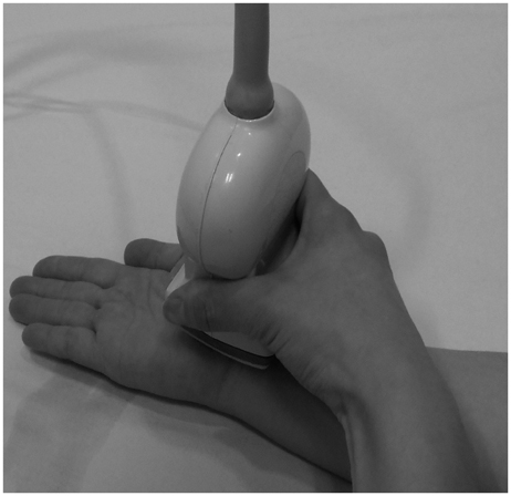
Fig. 1 3D ultrasonography of the median nerve in the carpal tunnel.
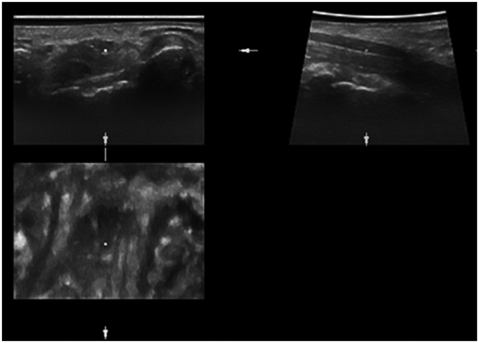
Fig. 2 Multiplanar display of a 3D ultrasonography volume showing the median nerve (dots) in three orthogonal orientations. The planes are interactive and permit a complete tracing of the median nerve from the distal radius to the proximal carpal tunnel.
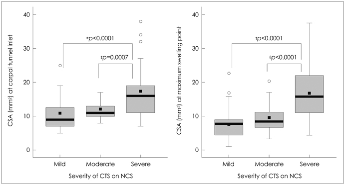
Fig. 3 Box plot showing the median nerve CSAs measured using 3D ultrasonography at the carpal tunnel inlet (left) and the maximal swelling point (right) in patients with CTS who were stratified according to the severity of their condition based on EDX (NCS). A gradual increase in CSA was observed with worsening CTS severity, with significant differences being apparent at both nerve sites between the severe- and mild-CTS groups (*p<0.0001 at the carpal tunnel inlet; †p<0.0001 at the maximum swelling point) and between the severe- and moderate-CTS groups (‡p=0.0007 at the carpal tunnel inlet; §p<0.0001 at the maximum swelling point). Bold black lines indicate medians and whiskers indicate error bars; ○, outliers; ■, mean values (10.5±4.6, 11.6±2.6, and 17.1±7.5 mm2 in the mild-, moderate-, and severe-CTS groups, respectively, at the carpal tunnel inlet; and 11.7±4.7, 13.0±3.4, and 19.7±7.2 mm2, respectively, at the maximum swelling point). CSAs: cross-sectional areas, CTS: carpal tunnel syndrome, EDX: electrodiagnostic, NCS: nerve conduction study.
Cited by 1 articles
-
Electrophysiologic and Ultrasonographic Assessment of Carpal Tunnel Syndrome in Wheelchair Basketball Athletes
Do Kyun Kim, Beom Suk Kim, Min Je Kim, Ki Hoon Kim, Byung Kyu Park, Dong Hwee Kim
Ann Rehabil Med. 2017;41(1):58-65. doi: 10.5535/arm.2017.41.1.58.
Reference
-
1. Atroshi I, Gummesson C, Johnsson R, Ornstein E, Ranstam J, Rosén I. Prevalence of carpal tunnel syndrome in a general population. JAMA. 1999; 282:153–158.
Article2. Phalen GS. The carpal-tunnel syndrome. Clinical evaluation of 598 hands. Clin Orthop Relat Res. 1972; 83:29–40.3. Lee D, van Holsbeeck MT, Janevski PK, Ganos DL, Ditmars DM, Darian VB. Diagnosis of carpal tunnel syndrome. Ultrasound versus electromyography. Radiol Clin North Am. 1999; 37:859–872. x4. Buchberger W, Schön G, Strasser K, Jungwirth W. High-resolution ultrasonography of the carpal tunnel. J Ultrasound Med. 1991; 10:531–537.
Article5. Wiesler ER, Chloros GD, Cartwright MS, Smith BP, Rushing J, Walker FO. The use of diagnostic ultrasound in carpal tunnel syndrome. J Hand Surg Am. 2006; 31:726–732.
Article6. Duncan I, Sullivan P, Lomas F. Sonography in the diagnosis of carpal tunnel syndrome. AJR Am J Roentgenol. 1999; 173:681–684.
Article7. Wong SM, Griffith JF, Hui AC, Tang A, Wong KS. Discriminatory sonographic criteria for the diagnosis of carpal tunnel syndrome. Arthritis Rheum. 2002; 46:1914–1921.
Article8. Koyuncuoglu HR, Kutluhan S, Yesildag A, Oyar O, Guler K, Ozden A. The value of ultrasonographic measurement in carpal tunnel syndrome in patients with negative electrodiagnostic tests. Eur J Radiol. 2005; 56:365–369.
Article9. Ziswiler HR, Reichenbach S, Vögelin E, Bachmann LM, Villiger PM, Jüni P. Diagnostic value of sonography in patients with suspected carpal tunnel syndrome: a prospective study. Arthritis Rheum. 2005; 52:304–311.
Article10. Padua L, Pazzaglia C, Caliandro P, Granata G, Foschini M, Briani C, et al. Carpal tunnel syndrome: ultrasound, neurophysiology, clinical and patient-oriented assessment. Clin Neurophysiol. 2008; 119:2064–2069.
Article11. Klauser AS, Halpern EJ, De Zordo T, Feuchtner GM, Arora R, Gruber J, et al. Carpal tunnel syndrome assessment with US: value of additional cross-sectional area measurements of the median nerve in patients versus healthy volunteers. Radiology. 2009; 250:171–177.
Article12. Dückelmann AM, Kalache KD. Three-dimensional ultrasound in evaluating the fetus. Prenat Diagn. 2010; 30:631–638.
Article13. Pyun SB, Kang CH, Yoon JS, Kwon HK, Kim JH, Chung KB, et al. Application of 3-dimensional ultrasonography in assessing carpal tunnel syndrome. J Ultrasound Med. 2011; 30:3–10.
Article14. Bayrak IK, Bayrak AO, Tilki HE, Nural MS, Sunter T. Ultrasonography in carpal tunnel syndrome: comparison with electrophysiological stage and motor unit number estimate. Muscle Nerve. 2007; 35:344–348.
Article15. Karadağ YS, Karadağ O, Ciçekli E, Oztürk S, Kiraz S, Ozbakir S, et al. Severity of Carpal tunnel syndrome assessed with high frequency ultrasonography. Rheumatol Int. 2010; 30:761–765.
Article16. Karmakar M, Li X, Li J, Sala-Blanch X, Hadzic A, Gin T. Three-dimensional/four-dimensional volumetric ultrasound imaging of the sciatic nerve. Reg Anesth Pain Med. 2012; 37:60–66.
Article17. Choquet O, Capdevila X. Case report: Three-dimensional high-resolution ultrasound-guided nerve blocks: a new panoramic vision of local anesthetic spread and perineural catheter tip location. Anesth Analg. 2013; 116:1176–1181.18. Girtler MT, Krasinski A, Dejaco C, Kitzler HH, Cui LG, Sherebrin S, et al. Feasibility of 3D ultrasound to evaluate upper extremity nerves. Ultraschall Med. 2013; 34:382–387.
Article19. Clendenen SR, Robards CB, Clendenen NJ, Freidenstein JE, Greengrass RA. Real-time 3-dimensional ultrasound-assisted infraclavicular brachial plexus catheter placement: implications of a new technology. Anesthesiol Res Pract. 2010; 2010:pii:208025.
Article20. Moran L, Perez M, Esteban A, Bellon J, Arranz B, del Cerro M. Sonographic measurement of cross-sectional area of the median nerve in the diagnosis of carpal tunnel syndrome: correlation with nerve conduction studies. J Clin Ultrasound. 2009; 37:125–131.
Article21. Lee CH, Kim TK, Yoon ES, Dhong ES. Correlation of high-resolution ultrasonographic findings with the clinical symptoms and electrodiagnostic data in carpal tunnel syndrome. Ann Plast Surg. 2005; 54:20–23.
Article22. Mondelli M, Filippou G, Gallo A, Frediani B. Diagnostic utility of ultrasonography versus nerve conduction studies in mild carpal tunnel syndrome. Arthritis Rheum. 2008; 59:357–366.
Article23. Moghtaderi A, Sanei-Sistani S, Sadoughi N, Hamed-Azimi H. Ultrasound evaluation of patients with moderate and severe carpal tunnel syndrome. Prague Med Rep. 2012; 113:23–32.
Article24. Ooi CC, Wong SK, Tan AB, Chin AY, Abu Bakar R, Goh SY, et al. Diagnostic criteria of carpal tunnel syndrome using high-resolution ultrasonography: correlation with nerve conduction studies. Skeletal Radiol. 2014; [Epub ahead of print].
Article
- Full Text Links
-
- Actions
-
Cited
- CITED
-
- Close
- Share
-
- Similar articles
-
- The Correlation Between Electrodiagnostic Results and Ultrasonographic Findings in the Severity of Carpal Tunnel Syndrome in Females
- Analysis of Sonographic Measurement by Anatomical Area in Carpal Tunnel Syndrome and Correlation the Measurement with Electrodiagnostic Study
- Ultrasonographic Study of Median Nerve after Carpal Tunnel Release
- Pressure Measurement in Carpal Tunnel Syndrome : Correlation with Electrodiagnostic and Ultrasonographic Findings
- The Correlation of High Resolution Ultrasonographic Findings with EMG and Clinical Symptoms
