Initial Experience with Magnetic Resonance-Guided Vacuum-Assisted Biopsy in Korean Women with Breast Cancer
- Affiliations
-
- 1Department of Radiology, Samsung Medical Center, Sungkyunkwan University School of Medicine, Seoul, Korea. bkhan@skku.edu
- KMID: 2286355
- DOI: http://doi.org/10.4048/jbc.2014.17.3.270
Abstract
- PURPOSE
The aim of this study is to describe our initial experience with magnetic resonance (MR)-guided biopsy and to determine the malignancy rate of additional lesions identified by MR only in Korean women with breast cancer.
METHODS
A retrospective review identified 22 consecutive patients with breast cancer who had undergone MR-guided vacuum-assisted biopsies (VAB) of MR-only identified lesions from May 2009 to October 2011.We evaluated the rate of compliance, the technical success for MR-guided VAB and the MR imaging findings of the target lesions. VAB histology was compared with surgical histology and follow-up imaging findings.
RESULTS
The biopsy recommendations for MR-only identified lesions were accepted in 46.8% (22/47) of patients. One of 22 procedures failed due to the target's posterior location. Among 21 MR-guided VAB procedures, the target lesions were considered as a mass in 12 cases and a nonmass enhancement in nine cases. VAB histology revealed malignancies in 14% (3/21) of cases, high-risk lesions in 24% (5/21) and benign lesions in 62% (13/21). Eleven cases (52%, 11/21) had a positive surgical correlation, and one of them was upgraded from atypical ductal hyperplasia to invasive ductal carcinoma. In the remaining 10 lesions, follow-up breast ultrasound and mammography were available (range, 15-44 months; mean, 32.1 months) and did not show suspicious lesions. The final malignancy rate was 19% (4/21).
CONCLUSION
MR-guided VAB for MR-only identified lesions yielded a 19% malignancy rate in Korean women with breast cancer. MR-guided VAB helps surgeons avoid an unnecessary wide excision or additional excisional biopsy.
MeSH Terms
Figure
-
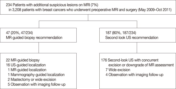
Figure 1 Flowchart for additional suspicious lesions on magnetic resonance imaging (MRI) in patients with breast cancer who underwent preoperative breast MRI and surgeries.US=ultrasound.
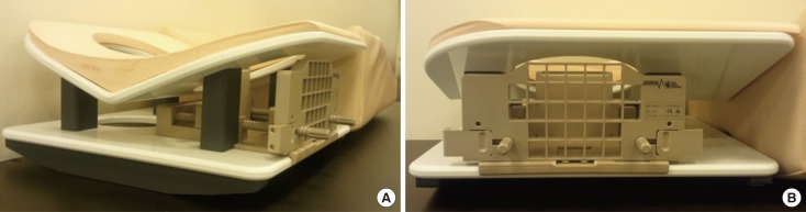
Figure 2 (A, B) Dedicated biopsy compression device and a commercially available compression grid-localizing system (Biopsy Positioning Device Model MR-BI-160, MRI Devices; GE Healthcare).
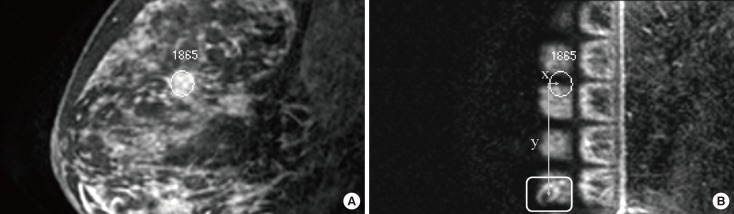
Figure 3 A method of determining the lesion location. (A, B) After reviewing the images on the console, a cursor was placed over the lesion (in a circle) and fiducial marker (in a rectangle) on the monitor. The differences in dorsal-ventral (x), cranial-caudal (y), and medial-lateral (z) direction coordinates of the lesion and fiducial marker were calculated on the basis of the spatial relationship between the lesion, vitamin E marker, and grid lines. The difference in z direction was calculated by a following formula: difference of imaging number of the lesion and fiducial marker×slice thickness of the magnetic resonance images.
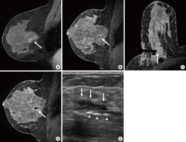
Figure 4 Images of a 49-year-old woman with diagnosed as ductal carcinoma in situ (DCIS) in the left breast. (A) Initial diagnostic contrast enhanced sagittal T1-weighted 3D turbo field-echo image shows segmental enhancing lesion without washout in the mid-outer region of the contralateral (right) breast (arrow). Mammograms and second-look ultrasound did not show clear relation with magnetic resonance (MR) imaging finding. (B) Sagittal prebiopsy MR image with the same sequence reveals the tip of inserted obturator at the targeted lesion (arrow). (C) Axial MR image confirms the exact location of the tip (arrow). (D) Sagittal MR image obtained after vacuum biopsy shows air at anterior to biopsy site and lesion disappearance (arrow). (E) Overt hematoma (arrows) and echogenic air collection (arrowheads) are seen on ultrasound (US). US-guided tattooing for the vacuum-assisted biopsy site was done before the surgery. MR-guided vacuum-assisted biopsy revealed DCIS. Operation after the biopsy confirmed DCIS.
Reference
-
1. Baum F, Fischer U, Vosshenrich R, Grabbe E. Classification of hypervascularized lesions in CE MR imaging of the breast. Eur Radiol. 2002; 12:1087–1092. PMID: 11976850.
Article2. Helbich TH. Contrast-enhanced magnetic resonance imaging of the breast. Eur J Radiol. 2000; 34:208–219. PMID: 10927162.
Article3. Peters NH, Borel Rinkes IH, Zuithoff NP, Mali WP, Moons KG, Peeters PH. Meta-analysis of MR imaging in the diagnosis of breast lesions. Radiology. 2008; 246:116–124. PMID: 18024435.
Article4. Fischer U, Kopka L, Grabbe E. Breast carcinoma: effect of preoperative contrast-enhanced MR imaging on the therapeutic approach. Radiology. 1999; 213:881–888. PMID: 10580970.
Article5. Orel SG, Schnall MD. MR imaging of the breast for the detection, diagnosis, and staging of breast cancer. Radiology. 2001; 220:13–30. PMID: 11425968.
Article6. Kuhl CK, Bieling HB, Gieseke J, Kreft BP, Sommer T, Lutterbey G, et al. Healthy premenopausal breast parenchyma in dynamic contrast-enhanced MR imaging of the breast: normal contrast medium enhancement and cyclical-phase dependency. Radiology. 1997; 203:137–144. PMID: 9122382.
Article7. Kuhl CK, Mielcareck P, Klaschik S, Leutner C, Wardelmann E, Gieseke J, et al. Dynamic breast MR imaging: are signal intensity time course data useful for differential diagnosis of enhancing lesions? Radiology. 1999; 211:101–110. PMID: 10189459.
Article8. Orel SG. Differentiating benign from malignant enhancing lesions identified at MR imaging of the breast: are time-signal intensity curves an accurate predictor? Radiology. 1999; 211:5–7. PMID: 10189447.
Article9. Nunes LW, Schnall MD, Orel SG. Update of breast MR imaging architectural interpretation model. Radiology. 2001; 219:484–494. PMID: 11323476.
Article10. Kuhl CK, Schmutzler RK, Leutner CC, Kempe A, Wardelmann E, Hocke A, et al. Breast MR imaging screening in 192 women proved or suspected to be carriers of a breast cancer susceptibility gene: preliminary results. Radiology. 2000; 215:267–279. PMID: 10751498.
Article11. Brown J, Smith RC, Lee CH. Incidental enhancing lesions found on MR imaging of the breast. AJR Am J Roentgenol. 2001; 176:1249–1254. PMID: 11312189.
Article12. Teifke A, Lehr HA, Vomweg TW, Hlawatsch A, Thelen M. Outcome analysis and rational management of enhancing lesions incidentally detected on contrast-enhanced MRI of the breast. AJR Am J Roentgenol. 2003; 181:655–662. PMID: 12933456.
Article13. Liberman L, Morris EA, Dershaw DD, Thornton CM, Van Zee KJ, Tan LK. Fast MRI-guided vacuum-assisted breast biopsy: initial experience. AJR Am J Roentgenol. 2003; 181:1283–1293. PMID: 14573421.14. Orel SG, Rosen M, Mies C, Schnall MD. MR imaging-guided 9-gauge vacuum-assisted core-needle breast biopsy: initial experience. Radiology. 2006; 238:54–61. PMID: 16304093.
Article15. Perlet C, Heywang-Kobrunner SH, Heinig A, Sittek H, Casselman J, Anderson I, et al. Magnetic resonance-guided, vacuum-assisted breast biopsy: results from a European multicenter study of 538 lesions. Cancer. 2006; 106:982–990. PMID: 16456807.16. Choi HY, Kim SM, Jang M, Yun BL, Kim SW, Kang E, et al. MRI-guided intervention for breast lesions using the freehand technique in a 3.0-T closed-bore MRI scanner: feasibility and initial results. Korean J Radiol. 2013; 14:171–178. PMID: 23482868.
Article17. LaTrenta LR, Menell JH, Morris EA, Abramson AF, Dershaw DD, Liberman L. Breast lesions detected with MR imaging: utility and histopathologic importance of identification with US. Radiology. 2003; 227:856–861. PMID: 12773685.
Article18. Liberman L, Morris EA, Lee MJ, Kaplan JB, LaTrenta LR, Menell JH, et al. Breast lesions detected on MR imaging: features and positive predictive value. AJR Am J Roentgenol. 2002; 179:171–178. PMID: 12076929.
Article19. Liberman L, Menell JH. Breast imaging reporting and data system (BI-RADS). Radiol Clin North Am. 2002; 40:409–430. PMID: 12117184.
Article20. Malhaire C, El Khoury C, Thibault F, Athanasiou A, Petrow P, Ollivier L, et al. Vacuum-assisted biopsies under MR guidance: results of 72 procedures. Eur Radiol. 2010; 20:1554–1562. PMID: 20119729.
Article21. Liberman L, Bracero N, Morris E, Thornton C, Dershaw DD. MRI-guided 9-gauge vacuum-assisted breast biopsy: initial clinical experience. AJR Am J Roentgenol. 2005; 185:183–193. PMID: 15972421.
Article22. Mahoney MC. Initial clinical experience with a new MRI vacuum-assisted breast biopsy device. J Magn Reson Imaging. 2008; 28:900–905. PMID: 18821610.
Article23. Lehman CD, Deperi ER, Peacock S, McDonough MD, Demartini WB, Shook J. Clinical experience with MRI-guided vacuum-assisted breast biopsy. AJR Am J Roentgenol. 2005; 184:1782–1787. PMID: 15908530.
Article24. Liberman L, Holland AE, Marjan D, Murray MP, Bartella L, Morris EA, et al. Underestimation of atypical ductal hyperplasia at MRI-guided 9-gauge vacuum-assisted breast biopsy. AJR Am J Roentgenol. 2007; 188:684–690. PMID: 17312054.
Article25. Han BK, Schnall MD, Orel SG, Rosen M. Outcome of MRI-guided breast biopsy. AJR Am J Roentgenol. 2008; 191:1798–1804. PMID: 19020252.
Article26. Tozaki M, Yamashiro N, Sakamoto M, Sakamoto N, Mizuuchi N, Fukuma E. Magnetic resonance-guided vacuum-assisted breast biopsy: results in 100 Japanese women. Jpn J Radiol. 2010; 28:527–533. PMID: 20799018.
Article27. Perlet C, Heinig A, Prat X, Casselman J, Baath L, Sittek H, et al. Multicenter study for the evaluation of a dedicated biopsy device for MR-guided vacuum biopsy of the breast. Eur Radiol. 2002; 12:1463–1470. PMID: 12042955.
Article28. Crystal P, Sadaf A, Bukhanov K, McCready D, O'Malley F, Helbich TH. High-risk lesions diagnosed at MRI-guided vacuum-assisted breast biopsy: can underestimation be predicted. Eur Radiol. 2011; 21:582–589. PMID: 20839000.
Article
- Full Text Links
-
- Actions
-
Cited
- CITED
-
- Close
- Share
-
- Similar articles
-
- Comparison of needle aspiration and vacuum-assisted biopsy in the ultrasound-guided drainage of lactational breast abscesses
- Treating Gynecomastia with Ultrasound-guided Vacuum-assisted Biopsy Device as a Cosmetic Method
- The Clinical Experience of an Ultrasound-guided Vacuum-assisted Resection (Mammotome) for Benign Breast Lesions through a Core Needle Biopsy
- The Usefulness of US-guided Vacuum-Assisted Breast Biopsy for Probably Benign Lesions
- Clinicopathologic Features of the Papillary Breast Lesions Diagnosed on Ultrasonography-guided Core Needle Biopsy
