Hyperosmotic Stimulus Down-regulates 1alpha, 25-dihydroxyvitamin D3-induced Osteoclastogenesis by Suppressing the RANKL Expression in a Co-culture System
- Affiliations
-
- 1Department of Oral Biology, Yonsei University, Seoul 120-752, Korea. lsi@yuhs.ac
- 2Department of Orthodontics, Yonsei University, Seoul 120-752, Korea.
- 3Oral Science Research Center, Yonsei University, Seoul 120-752, Korea.
- 4Brain Korea 21 Project of Dental Science, Yonsei University, Seoul 120-752, Korea.
- 5College of Dentistry, Yonsei University, Seoul 120-752, Korea.
- 6Department of Oral Microbiology, College of Dentistry, Chonnam National University, Gwangju 500-757, Korea.
- 7Department of Physiology, College of Medicine, Chungnam National University, Daejeon 305-764, Korea.
- KMID: 2285399
- DOI: http://doi.org/10.4196/kjpp.2010.14.3.169
Abstract
- The hyperosmotic stimulus is regarded as a mechanical factor for bone remodeling. However, whether the hyperosmotic stimulus affects 1alpha, 25-dihydroxyvitamin D3 (1alpha,25(OH)2D3)-induced osteoclastogenesis is not clear. In the present study, the effect of the hyperosmotic stimulus on 1alpha,25(OH)2D3-induced osteoclastogenesis was investigated in an osteoblast-preosteoclast co-culture system. Serial doses of sucrose were applied as a mechanical force. These hyperosmotic stimuli significantly evoked a reduced number of 1alpha,25(OH)2D3-induced tartrate-resistant acid phosphatase-positive multinucleated cells and 1alpha,25(OH)2D3-induced bone-resorbing pit area in a co-culture system. In osteoblastic cells, receptor activator of nuclear factor kappaB ligand (RANKL) and Runx2 expressions were down-regulated in response to 1alpha,25(OH)2D3. Knockdown of Runx2 inhibited 1alpha,25(OH)2D3-induced RANKL expression in osteoblastic cells. Finally, the hyperosmotic stimulus induced the overexpression of TonEBP in osteoblastic cells. These results suggest that hyperosmolarity leads to the down-regulation of 1alpha,25(OH)2D3-induced osteoclastogenesis, suppressing Runx2 and RANKL expression due to the TonEBP overexpression in osteoblastic cells.
Keyword
MeSH Terms
Figure
-

Fig. 1. Effect of hyperosmotic stimulus of sucrose on 1α,25(OH)2D3-induced osteoclast differentiation in a co-culture system. Mouse BMMs were co-cultured with calvarial osteoblastic cells in the presence of 1α,25(OH)2D3 and the indicated concentration of sucrose. (A) Preosteoclast cells underwent differentiation into TRAP-positive multinuclear cells in the presence of 1α,25(OH)2D3, and different concentrations of sucrose. (B) The number of TRAP-positive multinuclear cells was counted. (C) Cell viability was determined by a MTT assay. Results are presented as the mean values±SEM (n=4; ∗p<0.05, ∗∗p<0.001 versus 1α,25(OH)2D3 only). (D) Osteoclasts had bone resorbing activity on calcium phosphate apatite-coated plates. (E) Total areas of formation of pit resorption. Osteoclast differentiation was significantly inhibited as the increase in sucrose concentration. Bar indicates mean±SEM (n=5; ∗p<0.05, ∗∗p < 0.001 versus 1α,25(OH)2D3 only).
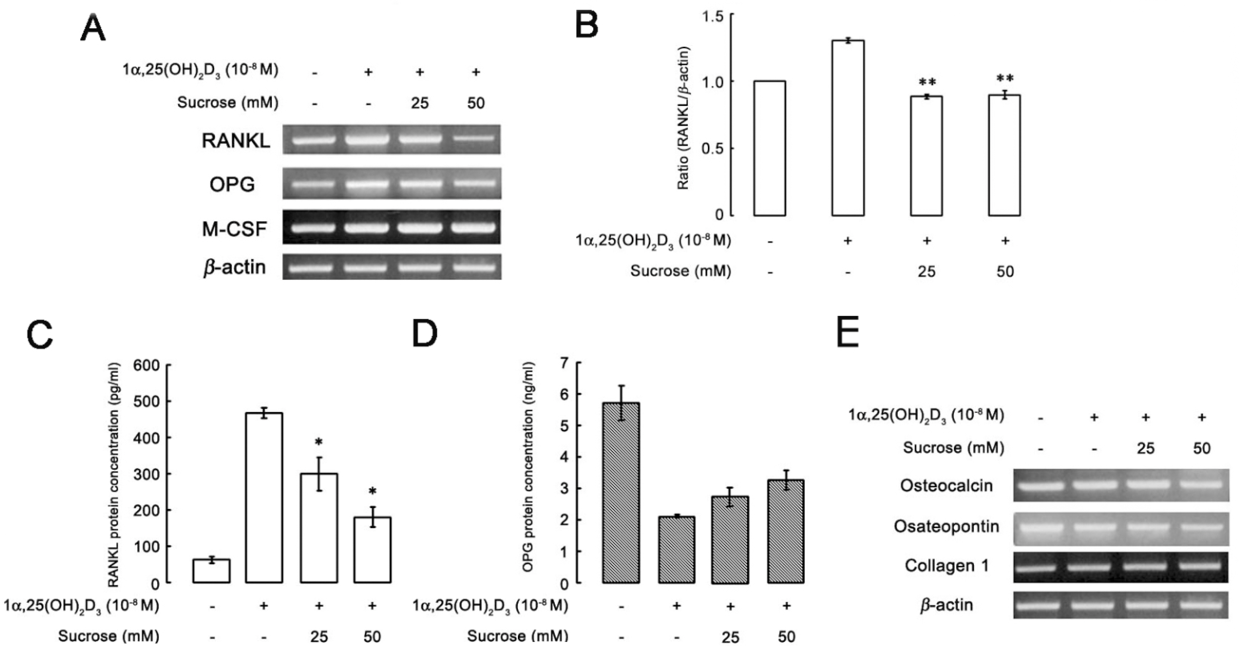
Fig. 2. Effect of hyperosmotic stimulus on the genes of osteoblast. Osteoblastic cells were cultured with 1α,25(OH)2D3 and the indicated sucrose concentration. (A) Total RNA was extracted, and then mRNA expression was analyzed by RT-PCR. (B) Bar graph shows the quantitative analysis of RANKL/β-actin mRNA. (C, D) For quantitative analysis, sRANKL and OPG productions were determined by ELISA. The bar graph shows the concentration level of RANKL and OPG protein. (E) Osteoblast marker genes were amplified by RT-PCR using RNA the primary osteoblastic cells that were exposed to the hyperosmotic stimulus. Bar represents the mean±SEM (n=4; ∗p<0.05 versus 1α,25(OH)2D3 only).
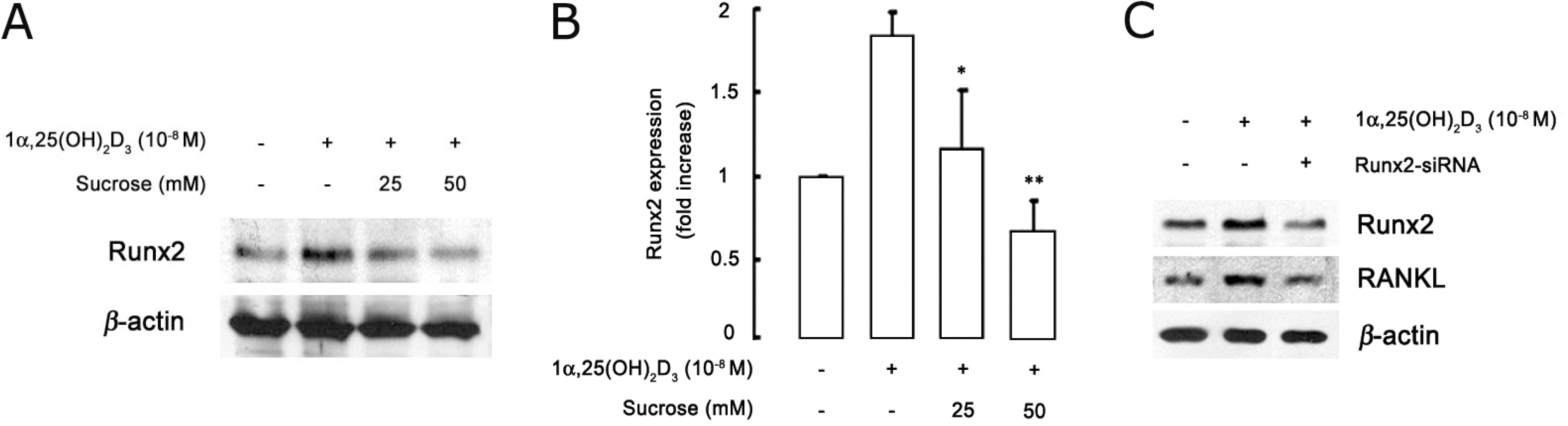
Fig. 3. Effects of hyperosmotic stimulus in 1α,25(OH)2D3-induced Runx2 expression. (A) Western blot analysis of Runx2 expression in osteoblastic cells stimulated with 1α, 25(OH)2D3 in the presence of indicated sucrose concentration. (B) Densitometric analysis of Runx2 and β-actin expression was used to normalize Runx2 data. (C) Western blot analysis of the levels of 1α,25(OH)2D3-induced RANKL and Runx2 expression in osteoblastic cells transfected with Runx2 siRNA and selected with G418-neomycine. Bar represents the mean±SEM (n=6; ∗p<0.05, ∗∗p < 0.001 versus 1α,25(OH)2D3 only).
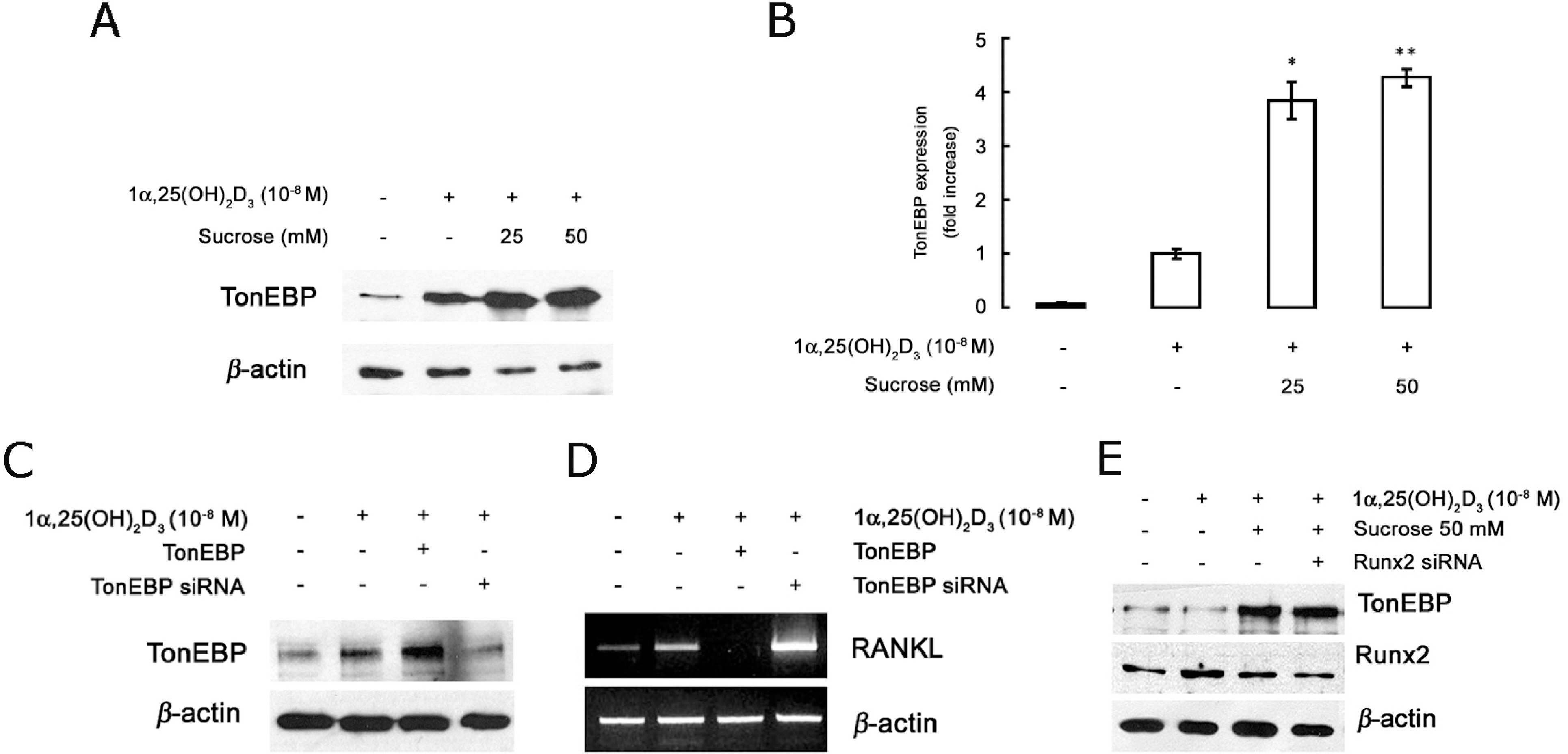
Fig. 4. Changes in the expression of TonEBP and Runx2 by hyperosmotic stimulus. Expression of TonEBP by hyperosmotic stress inhibits 1α,25(OH)2D3-induced RANKL expression in osteoblastic cells. (A) Western blot analysis of TonEBP expression in osteoblastic cells treated with 1α,25(OH)2D3 and 25 and 50 mM sucrose addition to the culture medium for 2 days. (B) TonEBP was stimulated by hyperosmotic stress caused by the addition of sucrose. (C) Western blot analysis of TonEBP expression levels in osteoblastic cells transfected with TonEBP (overexpression) or TonEBP siRNA. (D) The levels of RANKL expression were analyzed by RT-PCR using osteoblastic cells transfected with TonEBP siRNA and TonEBP in the presence or absence of 1α,25(OH)2D3. (E) The down-regulation in Runx2 expression by the stimulation of TonEBP (in response to the addition of 50 mM sucrose to the culture medium) in osteoblastic cells and TonEBP expression in osteoblastic cells transfected with Runx2 siRNA. Bar represents the mean±SEM (n=3; ∗p<0.05, ∗∗p<0.005 versus 1α,25(OH)2D3 only).
Reference
-
References
1. Teitelbaum SL. Bone resorption by osteoclasts. Science. 2000; 289:1504–1508.
Article2. Karsenty G, Wagner EF. Reaching a genetic and molecular understanding of skeletal development. Dev Cell. 2002; 2:389–406.
Article3. Suda T, Takahashi N, Martin TJ. Modulation of osteoclast differentiation. Endocr Rev. 1992; 13:66–80.
Article4. Takahashi M, Kushida K, Naitou K. The degree of osteoporosis in patients with vertebral fracture and patients with hip fracture: relationship to incidence of vertebral fracture. J Bone Miner Metab. 1999; 17:187–194.
Article5. Tsukii K, Shima N, Mochizuki S, Yamaguchi K, Kinosaki M, Yano K, Shibata O, Udagawa N, Yasuda H, Suda T, Higashio K. Osteoclast differentiation factor mediates an essential signal for bone resorption induced by 1 alpha, 25-dihydroxyvitamin D3, prostaglandin E2, or parathyroid hormone in the microenvironment of bone. Biochem Biophys Res Commun. 1998; 246:337–341.6. Boyle WJ, Simonet WS, Lacey DL. Osteoclast differentiation and activation. Nature. 2003; 423:337–342.
Article7. Teitelbaum SL, Ross FP. Genetic regulation of osteoclast development and function. Nat Rev Genet. 2003; 4:638–649.
Article8. Katagiri T, Takahashi N. Regulatory mechanisms of osteoblast and osteoclast differentiation. Oral Dis. 2002; 8:147–159.
Article9. Kitazawa S, Kitazawa R. RANK ligand is a prerequisite for cancer-associated osteolytic lesions. J Pathol. 2002; 198:228–236.
Article10. Takayanagi H, Iizuka H, Juji T, Nakagawa T, Yamamoto A, Miyazaki T, Koshihara Y, Oda H, Nakamura K, Tanaka S. Involvement of receptor activator of nuclear factor kappaB ligand/osteoclast differentiation factor in osteoclastogenesis from synoviocytes in rheumatoid arthritis. Arthritis Rheum. 2000; 43:259–269.11. Rodan GA, Martin TJ. Role of osteoblasts in hormonal control of bone resorption–a hypothesis. Calcif Tissue Int. 1981; 33:349–351.
Article12. Lacey DL, Timms E, Tan HL, Kelley MJ, Dunstan CR, Burgess T, Elliott R, Colombero A, Elliott G, Scully S, Hsu H, Sullivan J, Hawkins N, Davy E, Capparelli C, Eli A, Qian YX, Kaufman S, Sarosi I, Shalhoub V, Senaldi G, Guo J, Delaney J, Boyle WJ. Osteoprotegerin ligand is a cytokine that regulates osteoclast differentiation and activation. Cell. 1998; 93:165–176.
Article13. Burgess JR, David R, Greenaway TM, Parameswaran V, Shepherd JJ. Osteoporosis in multiple endocrine neoplasia type 1: severity, clinical significance, relationship to primary hyperparathyroidism, and response to parathyroidectomy. Arch Surg. 1999; 134:1119–1123.14. Kong YY, Feige U, Sarosi I, Bolon B, Tafuri A, Morony S, Capparelli C, Li J, Elliott R, McCabe S, Wong T, Campagnuolo G, Moran E, Bogoch ER, Van G, Nguyen LT, Ohashi PS, Lacey DL, Fish E, Boyle WJ, Penninger JM. Activated T cells regulate bone loss and joint destruction in adjuvant arthritis through osteoprotegerin ligand. Nature. 1999; 402:304–309.
Article15. Bao X, Clark CB, Frangos JA. Temporal gradient in shearinduced signaling pathway: involvement of MAP kinase, c-fos, and connexin43. Am J Physiolo Heart Circ Physiol. 2000; 278:H1598–H1605.16. Breen EC. Mechanical strain increases type I collagen expression in pulmonary fibroblasts in vitro. J Appl Physiol. 2000; 88:203–209.17. Chen NX, Ryder KD, Pavalko FM, Turner CH, Burr DB, Qiu J, Duncan RL. Ca2+ regulates fluid shear-induced cytoskeletal reorganization and gene expression in osteoblasts. Am J Physiol Cell Physiol. 2000; 278:C989–997.18. Klein-Nulend J, Helfrich MH, Sterck JG, MacPherson H, Joldersma M, Ralston SH, Semeins CM, Burger EH. Nitric oxide response to shear stress by human bone cells cultures is endothelial nitric oxide synthase dependent. Biochem and Biophys Res Commun. 1998; 250:108–114.19. Kreke MR, Huckle WR, Goldstein AS. Fluid flow stimulates expression of osteopontin and bone soialoprotein by bone marrow stromal cells in a temporally dependent manner. Bone. 2005; 36:1047–1055.20. Ingber DE. Tensegrity II. How structural networks influence cellular information processing networks. J Cell Sci. 2003; 116:1397–1408.
Article21. McGarry J, Klein-Nulend J, Prendergast PJ. The effect of cytoskeletal disruption on pulsatile fluid flow-induced nitric oxide and prostaglandin E(2) in ostocytes and ostoblast. Biochem Biophys Res Commun. 2005; 330:341–348.22. Yoshigi M, Hoffman LM, Jensen CC, Yost HJ, Beckerle MC. Mechanical force mobilizes zyxin from focal adhesions to actin filaments and regulates cytoskeletla reinforcement. J Cell Biol. 2005; 171:209–215.23. Miller SS, Wolf AM, Arnaud CD. Bone cells in culture: morphologic transformation by hormones. Science. 1976; 192:1340–1343.
Article24. Bowler WB, Buckley KA, Gartland A, Hipskind RA, Bilbe G, Gallagher JA. Extracellular nucleotide signaling: a mechanism for integrating local and systemic responses in the activation of bone remodeling. Bone. 2001; 28:507–512.
Article25. Marcus R. Normal and abnormal bone remodeling in man. Annu Rev Med. 1987; 38:129–141.
Article26. Choi BK, Ohk SH, Lee HJ, Kang JH, Jeong GJ, Yoo YJ. Effects of whole cell sonicates of Treponema lecithinolyticum on osteoclast differentiation. J Periodontol. 2001; 72:1172–1177.27. Mosmann T. Rapid colorimetric assay for cellular growth and survival: application to proliferation and cytotoxicity assays. J Immunol Methods. 1983; 65:55–63.
Article28. Tsai TT, Danielson KG, Guttapalli A, Oguz E, Albert TJ, Shapiro IM, Risbud MV. TonEBP/OREBP is a regulator of nucleus pulposus cell function and survival in the intervertebral disc. J Biol Chem. 2006; 281:25416–25424.
Article29. Takatsuna H, Asagiri M, Kubota T, Oka K, Osada T, Sugiyama C, Saito H, Aoki K, Ohya K, Takayanagi H, Umezawa K. Inhibition of RANKL-induced osteoclastogenesis by (–)-DHMEQ, a novel NF-kappaB inhibitor, through downregulation of NFATc1. J Bone Miner Res. 2005; 20:653–662.30. Kitazawa R, Kitazawa S, Maeda S. Promoter structure of mouse RANKL/TRANCE/OPGL/ODF gene. Biochim Biophys Acta. 1999; 1445:134–141.
Article31. Guilak F. Compression-induced changes in the shape and volume of the chondrocyte nucleus. J Biomech. 1995; 28:1529–1541.
Article32. Tsai TT, Danielson KG, Guttapalli A, Oguz E, Albert TJ, Shapiro IM, Risbud MV. TonEBP/OREBP is a regulator of nucleus pulposus cell function and survival in the intervertebral disc. J Biol Chem. 2006; 281:25416–24.
Article33. Tsuzuki T, Okabe K, Kajiya H, Habu T. Osmotic membrane stretch increases cytosolic Ca2+ and inhibits bone resorption activity in rat osteoclasts. Jpn J Physiol. 2000; 50:67–76.34. Zayzafoon M, Stell C, Irwin R, McCabe LR. Extracellular glucose influences osteoblast differentiation and c-jun expression. J Cell Biochem. 2000; 79:301–310.
Article35. Geoffroy V, Kneissel M, Fournier B, Boyde A, Matthias P. High bone resorption in adult aging transgenic mice overexpressing cbfa1/runx2 in cells of the osteoblastic lineage. Mol Cell Biol. 2002; 22:6222–6233.
Article36. Komori T, Yagi H, Nomura S, Yamaguchi A, Sasaki K, Deguchi K, Shimizu Y, Bronson RT, Gao YH, Inada M, Sato M, Okamoto R, Kitamura Y, Yoshiki S, Kishimoto T. Targeted disruption of Cbfa1 results in a complete lack of bone formation owing to maturational arrest of osteoblasts. Cell. 1997; 89:755–764.
Article37. Notoya M, Otsuka E, Yamaguchi A, Hagiwara H. Runx-2 is not essential for the vitamin D-regulated expression of RANKL and osteoprotegerin in osteoblastic cells. Biochem Biophys Res Commun. 2004; 324:655–660.
Article38. O'Brien CA, Kern B, Gubrij I, Karsenty G, Manolagas SC. Cbfa1 does not regulate RANKL gene activity in stromal/osteoblastic cells. Bone. 2002; 30:453–462.39. Sarkadi B, Parker JC. Activation of ion transport pathways by changes in cell volume. Biochim Biophys Acta. 1991; 1071:407–427.
Article
- Full Text Links
-
- Actions
-
Cited
- CITED
-
- Close
- Share
-
- Similar articles
-
- Hypoxia Inducible Factor-1alpha Directly Induces the Expression of Receptor Activator of Nuclear Factor-kappaB Ligand in MLO-Y4 Osteocytes
- Xylitol Down-Regulates 1alpha,25-Dihydroxy Vitamin D3-induced Osteoclastogenesis via in Part the Inhibition of RANKL Expression in Osteoblasts
- In Vitro Effects of 1,25-Dihydroxyvitamin D3 on the Production of Interleukin-1alpha by Ultraviolet B Irradiation in Cultured Human Keratinocyte Cell Line HaCaT Cells
- Caveolin-1 regulates osteoclast differentiation by suppressing cFms degradation
- The Effects of 1,25-Dihydroxyvitamin D3 on Expression of IGF-I Gene and Cellular Proliferation in MC3T3-E1 Cells
