Stimulatory Effects of Ginsan on the Proliferation and Viability of Mouse Spleen Cells
- Affiliations
-
- 1Laboratory of Veterinary Pharmacology, College of Veterinary Medicine, Jeju National University, Jeju 690-756, Korea. jooh@jejunu.ac.kr
- KMID: 2285393
- DOI: http://doi.org/10.4196/kjpp.2010.14.3.133
Abstract
- Ginsan is an acidic polysaccharide purified from Panax ginseng, a famous oriental herb. Although a variety of biological activities of ginsan have been studied, the effects of ginsan on spleen cells are not fully elucidated. We investigated the effect of ginsan on the viability and proliferation of spleen cells. Using Cell Counting Kit-8(R) solution and trypan blue solution, we found that ginsan significantly enhanced viability and proliferation. Multiple clusters, indicating proliferation, were observed in ginsan-treated spleen cells and, carboxyfluorescein succinimidyl ester and surface marker staining assay revealed that ginsan promoted proliferation from CD19+ B cells rather than CD4+ or CD8+ T cells. In addition, ginsan decreased the percentage of late apoptotic cells. Ginsan increased the surface expression of CD25 and CD69 as well as production of interleukin-2 from spleen cells, suggesting increased activation. Taken together, these results demonstrate that ginsan increases the viability and proliferation of spleen cells via multiple mechanisms, valuable information for broadening the use of ginsan in clinical and research settings.
Keyword
MeSH Terms
Figure
-

Fig. 1. Effect of ginsan on the proliferation/viability of spleen cells. The spleen cells were cultured at a concentration of 2×10⁵ cells/200 μl/ well in 96-well culture plates. (A) Proliferation assay using Cell Counting Kit-8® solution, in which spleen cells were treated with 0∼200 μg/ml ginsan for 4 days. (B) Trypan blue exclusion test, in which spleen cells were treated with 0∼100 μg/ml ginsan for 3 days. Data are mean±SD from three or four individual wells.
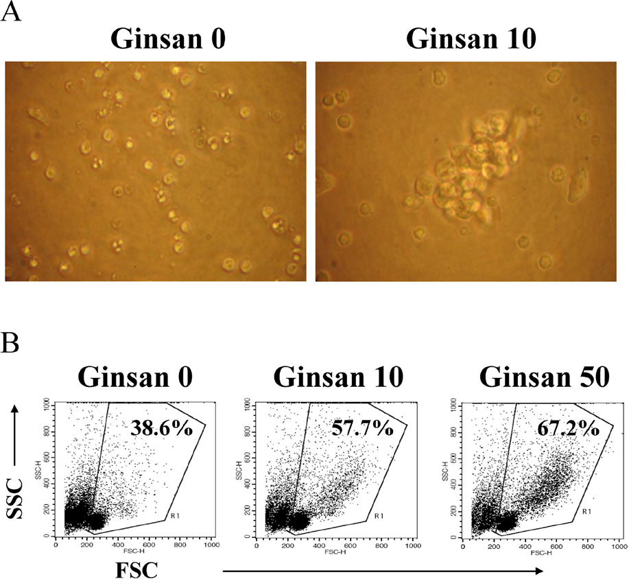
Fig. 2. Proliferating clusters in ginsan-treated spleen cells. Spleen cells were cultured as described in Fig. 1, and treated with medium alone, 10 μg/ml or 50 μg/ml ginsan for 3 days. (A) Cell morphology was observed using an inverted microscope and the image was obtained by a digital camera. (B) Cell size of the treated spleen cells was analyzed by flow cytometry.
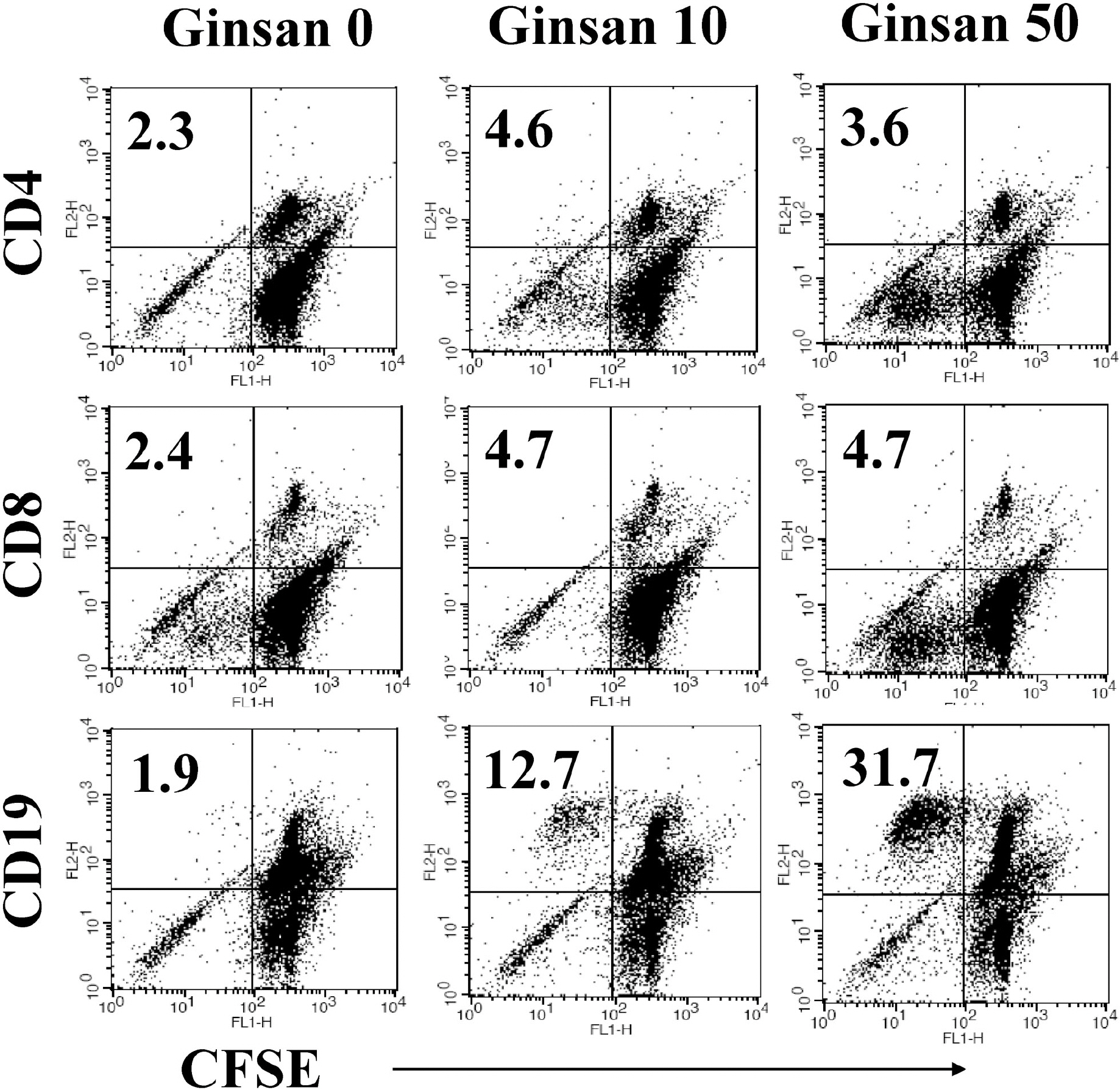
Fig. 3. Selective proliferation of CD19+ B lymphocytes by ginsan treatment. Spleen cells were stained with CFSE and cultured in 96-well culture plates with medium alone or 10 μg/ml or 50 μg/ml ginsan for 2 days. After harvesting, the cells were labeled with surface marker-specific antibodies. The numbers indicate the percentage of proliferating cells of a specific subset.

Fig. 4. Ginsan protects spleen cells against spontaneous cell death. Spleen cells were cultured at a concentration of 5×10⁶ cells/5 ml/well in 6-well culture plate. Following ginsan treatment, spleen cells were harvested and stained with annexin V-FITC/PI or rhodamine 123. The numbers indicate cell percentage and mean fluorescence intensity, respectively.
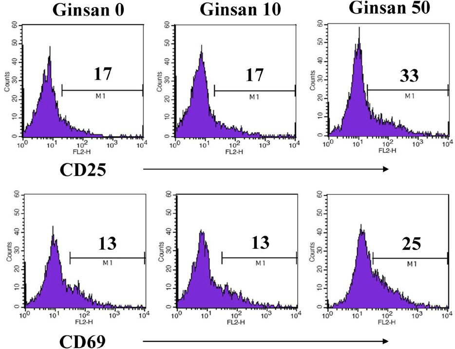
Fig. 5. Enhanced expression of activation markers on spleen cells treated with ginsan. Spleen cells were cultured and treated as described in Fig. 4. The number of histograms indicates the percentage of highly expressed cells.
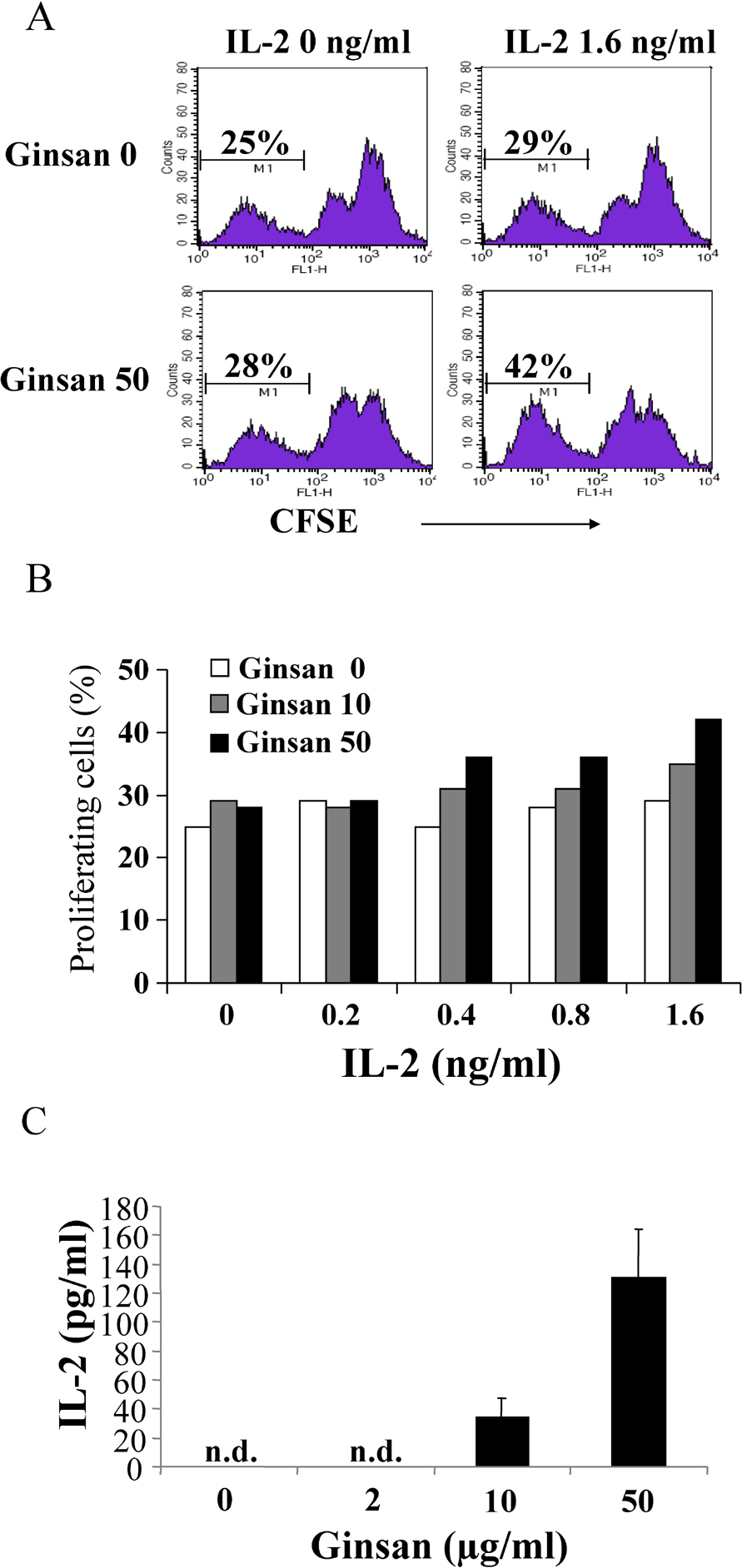
Fig. 6. IL-2 responsiveness of ginsan-activated spleen cells. (A, B) Spleen cells were cultured as described Fig. 4 and treated with ginsan for 2 days. The harvested cells were stained with CFSE and incubated in 24-well culture plates in the presence of IL-2 for 5 days. The percentage of low-intensity CFSE signals indicates proliferating cells. (C) Spleen cells were cultured as described in Fig. 1 and treated with the indicated concentrations of ginsan. After 3 days of treatment, the supernatants were collected and IL-2 and IL-4 were quantified. Data are mean±SD from four individual wells.
Cited by 1 articles
-
Immunostimulatory Effects of β-glucan Purified from
Paenibacillus polymyxa JB115 on Mouse Splenocytes
Ji-Mi Kim, Hong-Gu Joo
Korean J Physiol Pharmacol. 2012;16(4):225-230. doi: 10.4196/kjpp.2012.16.4.225.
Reference
-
References
1. Shim JY, Kim MH, Kim HD, Ahn JY, Yun YS, Song JY. Protective action of the immunomodulator ginsan against carbon tetrachloride-induced liver injury via control of oxidative stress and the inflammatory response. Toxicol Appl Pharmacol. 2010; 242:318–325.
Article2. Kim HJ, Kim MH, Byon YY, Park JW, Jee Y, Joo HG. Radio-protective effects of an acidic polysaccharide of Panax ginseng on bone marrow cells. J Vet Sci. 2007; 8:39–44.3. Shim JY, Han Y, Ahn JY, Yun YS, Song JY. Chemoprotective and adjuvant effects of immunomodulator ginsan in cyclophosphamide-treated normal and tumor bearing mice. Int J Immunopathol Pharmacol. 2007; 20:487–497.
Article4. Ahn JY, Choi IS, Shim JY, Yun EK, Yun YS, Jeong G, Song JY. The immunomodulator ginsan induces resistance to experimental sepsis by inhibiting Toll-like receptor-mediated inflammatory signals. Eur J Immunol. 2006; 36:37–45.
Article5. Kim MH, Byon YY, Ko EJ, Song JY, Yun YS, Shin T, Joo HG. Immunomodulatory activity of ginsan, a polysaccharide of Panax ginseng, on dendritic cells. Kor J Physiol Pharmacol. 2009; 13:169–173.6. Kim KH, Lee YS, Jung IS, Park SY, Chung HY, Lee IR, Yun YS. Acidic polysaccharide from Panax ginseng, ginsan, induces Th1 cell and macrophage cytokines and generates LAK cells in synergy with rIL-2. Planta Med. 1998; 64:110–115.7. Han SK, Song JY, Yun YS, Yi SY. Ginsan improved Th1 immune response inhibited by gamma radiation. Arch Pharm Res. 2005; 28:343–350.
Article8. Joo HG, Goedegebuure PS, Sadanaga N, Nagoshi M, von Bernstorff W, Eberlein TJ. Expression and function of galectin-3, a β-galactoside-binding protein in activated T lymphocytes. J Leukoc Biol. 2001; 69:555–564.9. Byon YY, Kim MH, Yoo ES, Hwang KK, Jee Y, Shin T, Joo HG. Radioprotective effects of fucoidan on bone marrow cells: improvement of the cell survival and immunoreactivity. J Vet Sci. 2008; 9:359–365.
Article10. Sugrue MM, Tatton WG. Mitochondrial membrane potential in aging cells. Biol Signals Recept. 2001; 10:176–188.
Article11. Hieronymus T, Blank N, Gruenke M, Winkler S, Haas JP, Kalden JR, Lorenz HM. CD 95-independent mechanisms of IL-2 deprivation-induced apoptosis in activated human lymphocytes. Cell Death Differ. 2000; 7:538–547.
Article12. Letai A. Growth factor withdrawal and apoptosis: the middle game. Mol Cell. 2006; 21:728–730.
Article13. Fleischer A, Duhamel M, Lopez-Fernandez LA, Muñoz M, Rebollo MP, Alvarez-Franco F, Rebollo A. Cascade of transcriptional induction and repression during IL-2 deprivation-induced apoptosis. Immunol Lett. 2007; 112:9–29.
Article14. Sancho D, Gómez M, Sánchez-Madrid F. CD69 is an immunore-gulatory molecule induced following activation. Trends Immunol. 2005; 6:136–140.
Article15. Rush JS, Hodgkin PD. B cells activated via CD40 and IL-4 undergo a division burst but require continued stimulation to maintain division, survival and differentiation. Eur J Immunol. 2001; 31:1150–1159.
Article16. Quigley M, Martinez J, Huang X, Yang Y. A critical role for direct TLR2-MyD88 signaling in CD8 T-cell clonal expansion and memory formation following vaccinia viral infection. Blood. 2009; 113:2256–2264.
Article17. Lee YS, Chung IS, Lee IR, Kim KH, Hong WS, Yun YS. Activation of multiple effector pathways of immune system by the antineoplastic immunostimulator acidic polysaccharide ginsan isolated from Panax ginseng. Anticancer Res. 1997; 17:323–331.18. Tangye SG, Hodgkin PD. Divide and conquer: the importance of cell division in regulating B-cell responses. Immunology. 2004; 112:509–520.
Article
- Full Text Links
-
- Actions
-
Cited
- CITED
-
- Close
- Share
-
- Similar articles
-
- Immunostimulatory effects of BCG-CWS on the proliferation and viability of mouse spleen cells
- Immunomodulatory Activity of Ginsan, a Polysaccharide of Panax Ginseng, on Dendritic Cells
- Induction of proliferation in resting B-cells by a factor released by activated mouse spleen cells
- Induction of apoptosis in mouse spleen cells by Ginsenoside Rp1
- Stimulatory Effect of IL-10 on Antitumor Cytolytic Activity of Murine Spleen Cells
