Accuracy of 3D white light scanning of abutment teeth impressions: evaluation of trueness and precision
- Affiliations
-
- 1Department of Dental Laboratory Science and Engineering, College of Health Science, Korea University, Seoul, Republic of Korea. kuc2842@korea.ac.kr
- 2Department of Public Health Sciences, Graduate School & BK21+ Program in Public Health Sciences, Korea University, Seoul, Republic of Korea.
- KMID: 2284720
- DOI: http://doi.org/10.4047/jap.2014.6.6.468
Abstract
- PURPOSE
This study aimed to evaluate the accuracy of digitizing dental impressions of abutment teeth using a white light scanner and to compare the findings among teeth types.
MATERIALS AND METHODS
To assess precision, impressions of the canine, premolar, and molar prepared to receive all-ceramic crowns were repeatedly scanned to obtain five sets of 3-D data (STL files). Point clouds were compared and error sizes were measured (n=10 per type). Next, to evaluate trueness, impressions of teeth were rotated by 10degrees-20degrees and scanned. The obtained data were compared with the first set of data for precision assessment, and the error sizes were measured (n=5 per type). The Kruskal-Wallis test was performed to evaluate precision and trueness among three teeth types, and post-hoc comparisons were performed using the Mann-Whitney U test with Bonferroni correction (alpha=.05).
RESULTS
Precision discrepancies for the canine, premolar, and molar were 3.7 microm, 3.2 microm, and 7.3 microm, respectively, indicating the poorest precision for the molar (P<.001). Trueness discrepancies for teeth types were 6.2 microm, 11.2 microm, and 21.8 microm, respectively, indicating the poorest trueness for the molar (P=.007).
CONCLUSION
In respect to accuracy the molar showed the largest discrepancies compared with the canine and premolar. Digitizing of dental impressions of abutment teeth using a white light scanner was assessed to be a highly accurate method and provided discrepancy values in a clinically acceptable range. Further study is needed to improve digitizing performance of white light scanning in axial wall.
Keyword
Figure
-
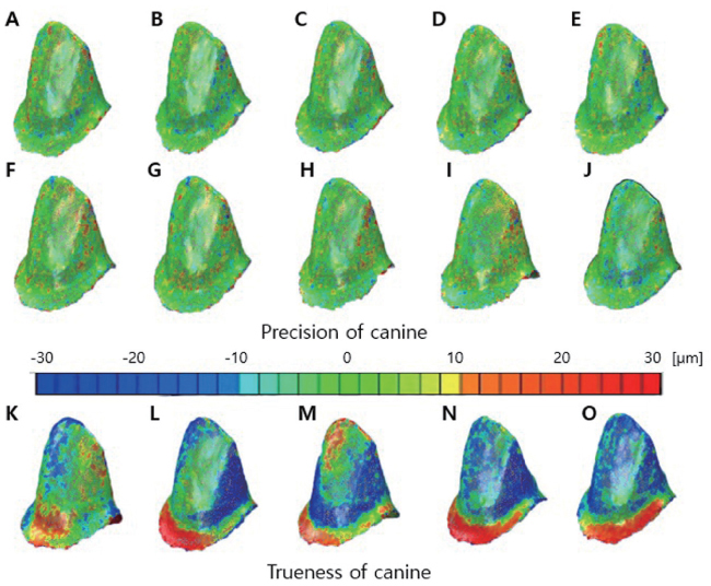
Fig. 1 Precision evaluation for the canine: Ten color-difference maps (A-J) of scans of the canine impression showing fit alignment of digital impression data compared among each other (C_pre1 through C_pre5). Trueness evaluation for the canine: Five color-difference maps (K-O) of scans of the canine impression showing fit alignment of digital impression data compared with the reference model data (C_tru1 to C_tru5). Green represents the exact fit, yellow to red represents a positive discrepancy, and turquoise to blue represents a negative discrepancy.
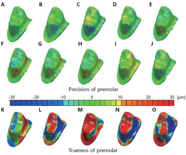
Fig. 2 Precision evaluation for the first premolar: Ten color-difference maps (A-J) of scans of the first premolar impression showing fit alignment of digital impression data compared among each other (P_pre1 through P_pre5). Trueness evaluation for the first premolar: Five color-difference maps (K-O) of scans of the first premolar impression showing fit alignment of digital impression data compared with the reference model (P_tru1 to P_tru5). Green represents the exact fit, yellow to red represents a positive discrepancy, and turquoise to blue represents a negative discrepancy.
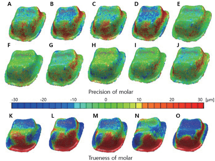
Fig. 3 Precision evaluation for the first molar: Ten color-difference maps (A-J) of scans of the first molar impression showing fit alignment of digital impression data compared among each other (M_pre1 through M_pre5). Trueness evaluation for the first molar: Five color-difference maps (K-O) of scans of the first molar impression showing fit alignment of digital impression data compared with the reference model (M_tru1 to M_tru5). Green represents the exact fit, yellow or red represents a positive discrepancy, and turquoise to blue represents a negative discrepancy.
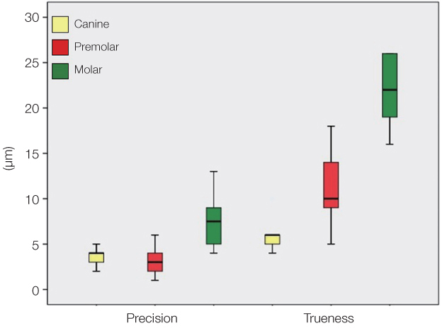
Fig. 4 Boxplot of discrepancies (precision and trueness) for the digitized impressions of the three types of abutment teeth obtained using a white light scanner: Precision (n=10 per type), trueness (n=5 per type).
Cited by 2 articles
-
Trueness and precision of scanning abutment impressions and stone models according to dental CAD/CAM evaluation standards
Jin-Hun Jeon, Seong-Sig Hwang, Ji-Hwan Kim, Woong-Chul Kim
J Adv Prosthodont. 2018;10(5):335-339. doi: 10.4047/jap.2018.10.5.335.Evaluation of the reproducibility of various abutments using a blue light model scanner
Dong-Yeon Kim, Kyung-Eun Lee, Jin-Hun Jeon, Ji-Hwan Kim, Woong-Chul Kim
J Adv Prosthodont. 2018;10(4):328-334. doi: 10.4047/jap.2018.10.4.328.
Reference
-
1. Naidu D, Freer TJ. Validity, reliability, and reproducibility of the iOC intraoral scanner: a comparison of tooth widths and Bolton ratios. Am J Orthod Dentofacial Orthop. 2013; 144:304–310.2. Flügge TV, Schlager S, Nelson K, Nahles S, Metzger MC. Precision of intraoral digital dental impressions with iTero and extraoral digitization with the iTero and a model scanner. Am J Orthod Dentofacial Orthop. 2013; 144:471–478.3. Persson AS, Odén A, Andersson M, Sandborgh-Englund G. Digitization of simulated clinical dental impressions: virtual three-dimensional analysis of exactness. Dent Mater. 2009; 25:929–936.4. Quaas S, Rudolph H, Luthardt RG. Direct mechanical data acquisition of dental impressions for the manufacturing of CAD/CAM restorations. J Dent. 2007; 35:903–908.5. Persson A, Andersson M, Oden A, Sandborgh-Englund G. A three-dimensional evaluation of a laser scanner and a touchprobe scanner. J Prosthet Dent. 2006; 95:194–200.6. Persson AS, Odén A, Andersson M, Sandborgh-Englund G. Digitization of simulated clinical dental impressions: virtual three-dimensional analysis of exactness. Dent Mater. 2009; 25:929–936.7. Schaefer O, Watts DC, Sigusch BW, Kuepper H, Guentsch A. Marginal and internal fit of pressed lithium disilicate partial crowns in vitro: a three-dimensional analysis of accuracy and reproducibility. Dent Mater. 2012; 28:320–326.8. Chang M, Park SC. Automated scanning of dental impressions. Comput Aided Des. 2009; 41:404–411.9. Brosky ME, Pesun IJ, Lowder PD, Delong R, Hodges JS. Laser digitization of casts to determine the effect of tray selection and cast formation technique on accuracy. J Prosthet Dent. 2002; 87:204–209.10. Khardekar R, Burton G, McMains S. Finding feasible mold parting directions using graphics hardware. Comput Aided Des. 2006; 38:327–341.11. Priyadarshi AK, Gupta SK. Geometric algorithms for automated design of multi-piece permanent molds. Comput Aided Des. 2004; 36:241–260.12. ISO 5725-1:2003. Accuracy (trueness and precision) of measurement methods and results? Part 1: General principles and definitions. Geneva; Swizterland: ISO;2003.13. Hoyos A, Soderholm KJ. Influence of tray rigidity and impression technique on accuracy of polyvinyl siloxane impressions. Int J Prosthodont. 2011; 24:49–54.14. Ziegler M. Digital impression taking with reproducibly high precision. Int J Comput Dent. 2009; 12:159–163.15. Chandran DT, Jagger DC, Jagger RG, Barbour ME. Two- and three-dimensional accuracy of dental impression materials: effects of storage time and moisture contamination. Biomed Mater Eng. 2010; 20:243–249.16. Wöstmann B, Rehmann P, Balkenhol M. Accuracy of impressions obtained with dual-arch trays. Int J Prosthodont. 2009; 22:158–160.17. Alikhasi M, Monzavi A, Bassir SH, Naini RB, Khosronedjad N, Keshavarz S. A comparison of precision of fit, rotational freedom, and torque loss with copy-milled zirconia and prefabricated titanium abutments. Int J Oral Maxillofac Implants. 2013; 28:996–1002.18. Webber B, McDonald A, Knowles J. An in vitro study of the compressive load at fracture of Procera AllCeram crowns with varying thickness of veneer porcelain. J Prosthet Dent. 2003; 89:154–160.19. Quaas S, Rudolph H, Luthardt RG. Direct mechanical data acquisition of dental impressions for the manufacturing of CAD/CAM restorations. J Dent. 2007; 35:903–908.20. Luthardt RG, Loos R, Quaas S. Accuracy of intraoral data acquisition in comparison to the conventional impression. Int J Comput Dent. 2005; 8:283–294.21. Kenyon BJ, Hagge MS, Leknius C, Daniels WC, Weed ST. Dimensional accuracy of 7 die materials. J Prosthodont. 2005; 14:25–31.22. DeLong R, Heinzen M, Hodges JS, Ko CC, Douglas WH. Accuracy of a system for creating 3D computer models of dental arches. J Dent Res. 2003; 82:438–442.23. Rudolph H, Luthardt RG, Walter MH. Computer-aided analysis of the influence of digitizing and surfacing on the accuracy in dental CAD/CAM technology. Comput Biol Med. 2007; 37:579–587.24. Jeon JH, Lee KT, Kim HY, Kim JH, Kim WC. White light scanner-based repeatability of 3-dimensional digitizing of silicon rubber abutment teeth impressions. J Adv Prosthodont. 2013; 5:452–456.25. Persson M, Andersson M, Bergman B. The accuracy of a high-precision digitizer for CAD/CAM of crowns. J Prosthet Dent. 1995; 74:223–229.26. Moldovan O, Luthardt RG, Corcodel N, Rudolph H. Three-dimensional fit of CAD/CAM-made zirconia copings. Dent Mater. 2011; 27:1273–1278.27. Persson AS, Andersson M, Odén A, Sandborgh-Englund G. Computer aided analysis of digitized dental stone replicas by dental CAD/CAM technology. Dent Mater. 2008; 24:1123–1130.
- Full Text Links
-
- Actions
-
Cited
- CITED
-
- Close
- Share
-
- Similar articles
-
- Trueness and precision of scanning abutment impressions and stone models according to dental CAD/CAM evaluation standards
- Comparing the accuracy of six intraoral scanners on prepared teeth and effect of scanning sequence
- White light scanner-based repeatability of 3-dimensional digitizing of silicon rubber abutment teeth impressions
- Accuracy of casts produced from conventional and digital workflows: A qualitative and quantitative analyses
- A novel reference model for dental scanning system evaluation: analysis of five intraoral scanners
