New Parametric Imaging Method with Fluorescein Angiograms for Detecting Areas of Capillary Nonperfusion
- Affiliations
-
- 1Biomedical Engineering Branch, Division of Convergence Technology, Research Institute, National Cancer Center, Goyang, Korea. kimkg@ncc.re.kr
- 2Department of Plasma Bio Display, Kwangwoon Universiy, Seoul, Korea.
- 3Department of Ophthalmology, Seoul National University Bundang Hospital, Seoul National University College of Medicine, Seongnam, Korea.
- KMID: 2284582
- DOI: http://doi.org/10.4258/hir.2014.20.3.191
Abstract
OBJECTIVES
Fluorescein angiography (FAG) is currently the most useful diagnostic modality for examining retinal circulation, and it is frequently used for the evaluation of patients with diabetic retinopathy, occlusive diseases, such as retinal venous and arterial occlusions, and wet macular degeneration. This paper presents a method for objectively evaluating retinal circulation by quantifying circulation-related parameters.
METHODS
This method allows the semiautomatic preprocessing and registering of FAG images. The arterial input function is estimated from the registered set of FAG images using gamma-variate fitting. Then, the parameters can be computed by deconvolution on the basis of truncated singular value decomposition, and they can finally be presented as parametric color images in a combination of three colors, red, green, and blue.
RESULTS
After the estimation of arterial input function, the parameters of relative blood flow and mean transit time were computed using deconvolution analysis based on truncated singular value decomposition.
CONCLUSIONS
The parametric color image is helpful to interpret the status of retinal blood circulation and provides quantitative data on retina ischemia without interobserver variability. This system easily provides the status of retinal blood circulation both qualitatively and quantitatively. It also helps to standardize FAG interpretation and may contribute to network-based telemedicine systems in the future.
Keyword
MeSH Terms
Figure
-

Figure 1 Flow diagram for image registration. CLAHE: contrast-limited adaptive histogram equalization.
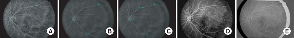
Figure 2 The result of image registration: (A) reference image, (B) moving image, (C) registered moving image, (D/E) difference image before/after registration.
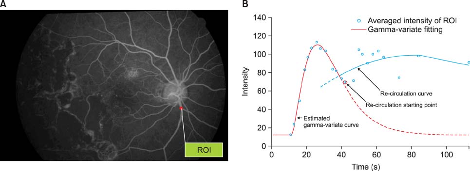
Figure 3 (A) A red region of interest (ROI) at the artery area. (B) The result of gamma-variate curve fitting for estimation estimating of the arterial input function.
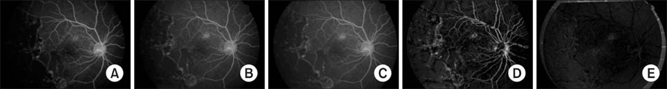
Figure 4 (A) Reference image, (B) moving image before registration, (C) moving image after registration, (D) difference image between (A) and (B), and (E) difference image between (A) and (C).
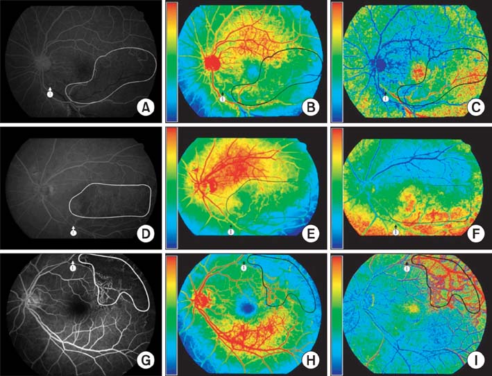
Figure 5 Fluorescein angiogram images (A, D, G), parametric images of blood flow (B, E, H) and mean transit time (C, F, I).
Reference
-
1. Koyama T, Matsuo N, Shimizu K, Mihara M, Tsuchida Y, Wolf S, et al. Retinal circulation times in quantitative fluorescein angiography. Graefes Arch Clin Exp Ophthalmol. 1990; 228(5):442–446.
Article2. Hickam JB, Frayser R. Aphotographic method for measuring the mean retinal circulation time using fluorescein. Invest Ophthalmol. 1965; 4(5):876–884.3. Sperber GO, Alm A. Retinal mean transit time determined with an impulse-response analysis from video fluorescein angiograms. Acta Ophthalmol Scand. 1997; 75(5):532–536.
Article4. Wintermark M, Sesay M, Barbier E, Borbely K, Dillon WP, Eastwood JD, et al. Comparative overview of brain perfusion imaging techniques. Stroke. 2005; 36(9):e83–e99.
Article5. Ostergaard L. Principles of cerebral perfusion imaging by bolus tracking. J Magn Reson Imaging. 2005; 22(6):710–717.
Article6. Ferreira RM, Lev MH, Goldmakher GV, Kamalian S, Schaefer PW, Furie KL, et al. Arterial input function placement for accurate CT perfusion map construction in acute stroke. AJR Am J Roentgenol. 2010; 194(5):1330–1336.
Article7. Huang LK, Wang MJ. Image thresholding by minimizing the measures of fuzziness. Pattern Recognit. 1995; 28(1):41–51.
Article8. Chanwimaluang T, Fan G, Fransen SR. Hybrid retinal image registration. IEEE Trans Inf Technol Biomed. 2006; 10(1):129–142.
Article9. Goatman KA, Manivannan A, Hipwell JH, Sharp PF, Lois N, Forrester JV. Automatic registration and averaging of ophthalmic autofluorescence images. In : Proceedings of the 5th Medical Image Understanding and Analysis; 2001 Jul 16-17; Birmingham, UK. p. 157–160.10. Dreo J, Nunes JC, Siarry P. Robust rigid registration of retinal angiograms through optimization. Comput Med Imaging Graph. 2006; 30(8):453–463.
Article11. Ryan N, Heneghan C, de Chazal P. Registration of digital retinal images using landmark correspondence by expectation maximization. Image Vis Comput. 2004; 22(11):883–898.
Article12. Jeong CB, Kim KG, Kim TS, Kim SK. Comparison of image enhancement methods for the effective diagnosis in successive whole-body bone scans. J Digit Imaging. 2011; 24(3):424–436.
Article13. Bisdas S, Medov L, Baghi M, Konstantinou GN, Wagenblast J, Thng CH, et al. A comparison of tumour perfusion assessed by deconvolution-based analysis of dynamic contrast-enhanced CT and MR imaging in patients with squamous cell carcinoma of the upper aerodigestive tract. Eur Radiol. 2008; 18(4):843–850.
Article14. He L, Orten BB, Do S, Karl WC, Kambadakone A, Sahani DV, et al. Spatio-temporal deconvolution of perfusion CT data in rectal tumor patients. In : Proceedings of the IEEE International Symposium on Biomedical Imaging: From Nano to Macro; 2009 Jun 28 - Jul 1; Boston, MA. p. 1231–1234.15. Chrastek R, Wolf M, Donath K, Niemann H, Paulus D, Hothorn T, et al. Automated segmentation of the optic nerve head for diagnosis of glaucoma. Med Image Anal. 2005; 9(4):297–314.
Article16. Fadzil MH, Nugroho HA, Nugroho H, Iznita IL. Contrast enhancement of retinal vasculature in digital fundus image. In : Proceedings of the International Conference on Digital Image Processing; 2009 Mar 7-9; Thailand. p. 137–141.17. Hani AF, Nugroho HA. Model-based retinal vasculature enhancement in digital fundus image using independent component analysis. In : Proceedings of the IEEE Symposium on Industrial Electronics & Applications; 2009 Oct 4-6; Kuala Lumpur, Malaysia. p. 160–164.18. Hipwell JH, Manivannan A, Vieira P, Sharp PF, Forrester JV. Quantifying changes in retinal circulation: the generation of parametric images from fluorescein angiograms. Physiol Meas. 1998; 19(2):165–180.
Article19. Hua C, Brener N, Thompson H, Iyengar SS, Zhengmao Y. Automated control point detection, registration, and fusion of fuzzy retinal vasculature images. In : Proceedings of the IEEE International Conference on Fuzzy Systems; 2008 Jun 1-6; Hong Kong, China. p. 2386–2391.
- Full Text Links
-
- Actions
-
Cited
- CITED
-
- Close
- Share
-
- Similar articles
-
- Bone Marrow Transplantation Retinopathy in a Patient with Acute Lymophocytic Leukemia Following Bone Marrow Transplantation
- The Relationship between the Capillary Nonperfusion Area and the Retinal Nerve Fiber Layer Defects in Branch Retinal Vein Occlusion Patients
- A Case of Purtscher's Retinopathy Responsive to High-dose Steroid Therapy
- Collateral Circulation in the Branch Retinal Vein Occlusion
- Different Modality of Treatment in the Neovascularization of Uveitis According to Nonperfusion Area
