Diagnosis and Treatment of Moyamoya Disease
- Affiliations
-
- 1Department of Neurosurgery, Ewha Womans University School of Medicine, Seoul, Korea. drekseo@ewha.ac.kr
- KMID: 2284010
- DOI: http://doi.org/10.12771/emj.2013.36.1.9
Abstract
- Moyamoya disease is a cerebrovascular disease of unknown etiology, which is characterized by bilateral stenosis or occlusion at terminal portion of internal carotid artery and at proximal portion of anterior cerebral artery and/or middle cerebral artery and abnormal vascular network in the vicinity of the arterial occlusions. It occurs frequently in Asian countries, particularly in Korea and Japan, but is rare in Western countries. To establish the etiology of moyamoya disease, much about the pathology from autopsies, factors involved in its pathogenesis, and its genetics have been studied. It may occur at any age from childhood to adulthood and in general, initial manifestation is cerebral ischemic symptoms in children and intracranial hemorrhage symptoms in adults. Because it progress and cause recurrent stroke, early diagnosis and proper management has been recognized. Cerebral angiography is essential for definitive diagnosis and treatment plan. Magnetic resonance imaging/magnetic resonance angiography is useful for diagnosis and follow-up tools after revascularization. Evaluation of the cerebral hemodynamics by single photon emission computed tomography and positron emission tomography is useful for diagnosis and assessment of the severity of cerebral ischemia in moyamoya patients. Surgical revascularization is effective for moyamoya disease manifesting as ischemic symptoms, to prevent further ischemia and infarction. In hemorrhagic type moyamoya disease, revascularization can be considered. Direct bypass, indirect synangiosis and combined methods are used. Outcomes of revascularization are excellent in preventing transient ischemic attacks in most patients.
MeSH Terms
-
Adult
Angiography
Anterior Cerebral Artery
Asian Continental Ancestry Group
Autopsy
Brain Ischemia
Carotid Artery, Internal
Cerebral Angiography
Cerebral Hemorrhage
Child
Constriction, Pathologic
Early Diagnosis
Follow-Up Studies
Hemodynamics
Humans
Infarction
Intracranial Hemorrhages
Ischemia
Ischemic Attack, Transient
Japan
Korea
Magnetic Resonance Spectroscopy
Middle Cerebral Artery
Moyamoya Disease
Positron-Emission Tomography
Stroke
Tomography, Emission-Computed, Single-Photon
Figure
-

Fig. 1 A left carotid angiography (A, anteroposterior view; B, lateral view) shows severe stenosis in terminal portion of internal carotid artery and proximal middle cerebral artery and occlusion of proximal portion of anterior cerebral artery and demonstrates basal collateral vessels. A right carotid angiography (C, anteroposterior view; D, lateral view) demonstrates stenosis of proximal middle cerebral artery and occlusion in proximal portion of anterior cerebral artery.

Fig. 2 Initial angiography (A) shows mild stenosis in terminal portion of internal carotid artery and well visualized middle cerebral artery. After 2 years, angiography (B) demonstrate that middle cerebral artery is not visualized and basal there are collateral vessels (arrow head) from posterior cerebral artery. Posterior cerebral artery is occluded and collateral vessel is disappeared after 6 months later (C).
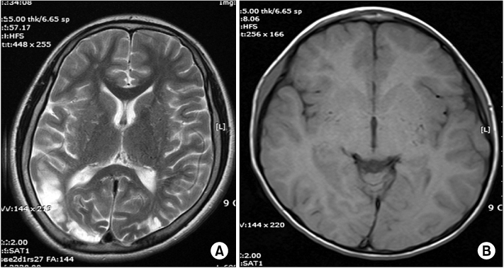
Fig. 3 A T2 weighted axial magnetic resonance image (A) shows cerebral infarction in right temporo-occipital areas. An axial T1 weighted image (B) demonstrates multiple signal voids of the hypertrophied moyamoya collateral in the basal ganglia.

Fig. 4 Basal single photon emission computed tomography (SPECT) (A) and acetazolamide SPECT (B) show decreased blood flow and reserve capacity in left frontal lobe.
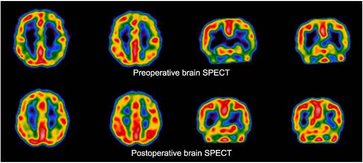
Fig. 5 After revascularization, cerebral blood flow is increased in left frontal lobe.
Reference
-
1. Suzuki J, Takaku A. Cerebrovascular "moyamoya" disease. Disease showing abnormal net-like vessels in base of brain. Arch Neurol. 1969. 20:288–299.2. Natori Y, Ikezaki K, Matsushima T, Fukui M. 'Angiographic moyamoya' its definition, classification, and therapy. Clin Neurol Neurosurg. 1997. 99:Suppl 2. S168–S172.3. Czartoski T, Hallam D, Lacy JM, Chun MR, Becker K. Postinfectious vasculopathy with evolution to moyamoya syndrome. J Neurol Neurosurg Psychiatry. 2005. 76:256–259.4. Suzuki J, Kodama N. Moyamoya disease: a review. Stroke. 1983. 14:104–109.5. Kuroda S, Ishikawa T, Houkin K, Nanba R, Hokari M, Iwasaki Y. Incidence and clinical features of disease progression in adult moyamoya disease. Stroke. 2005. 36:2148–2153.6. Nakashima H, Meguro T, Kawada S, Hirotsune N, Ohmoto T. Long-term results of surgically treated moyamoya disease. Clin Neurol Neurosurg. 1997. 99:Suppl 2. S156–S161.7. Okada Y, Shima T, Nishida M, Yamane K, Yamada T, Yamanaka C. Effectiveness of superficial temporal artery-middle cerebral artery anastomosis in adult moyamoya disease: cerebral hemodynamics and clinical course in ischemic and hemorrhagic varieties. Stroke. 1998. 29:625–630.8. Goto Y, Yonekawa Y. Worldwide distribution of moyamoya disease. Neurol Med Chir (Tokyo). 1992. 32:883–886.9. Wakai K, Tamakoshi A, Ikezaki K, Fukui M, Kawamura T, Aoki R, et al. Epidemiological features of moyamoya disease in Japan: findings from a nationwide survey. Clin Neurol Neurosurg. 1997. 99:Suppl 2. S1–S5.10. Ohki K, Hoshino H, Nogawa S, Yamaguchi K. Ministry of Health, Labour and Welfare. 2006 Databases evaluation by the Research Committee on Moyamoya Disease (Spontaneous occlusion of the circle of Willis). Research on spontaneous occlusion of the circle of Willis. 2007. Tokyo: Ministry of Health, Labour and Welfare;19–25.11. Ikezaki K. American Association of Neurological Surgeons. Clinical manifestations: epidemiology, symptoms and signs, laboratory findings. Moyamoya disease. 2001. Rolling Meadows (IL): American Association of Neurological Surgeons;43–54.12. Yamaguchi K, Nogawa S, Fukuuchi Y. National survey on spontaneous occlusion of the circle of Willis (moyamoya disease). Shinkei Naika. 2001. 54:319–327.13. Baba T, Houkin K, Kuroda S. Novel epidemiological features of moyamoya disease. J Neurol Neurosurg Psychiatry. 2008. 79:900–904.14. Fukui M, Kono S, Sueishi K, Ikezaki K. Moyamoya disease. Neuropathology. 2000. 20:Suppl. S61–S64.15. Malek AM, Connors S, Robertson RL, Folkman J, Scott RM. Elevation of cerebrospinal fluid levels of basic fibroblast growth factor in moyamoya and central nervous system disorders. Pediatr Neurosurg. 1997. 27:182–189.16. Hojo M, Hoshimaru M, Miyamoto S, Taki W, Nagata I, Asahi M, et al. Role of transforming growth factor-beta1 in the pathogenesis of moyamoya disease. J Neurosurg. 1998. 89:623–629.17. Mineharu Y, Takenaka K, Yamakawa H, Inoue K, Ikeda H, Kikuta KI, et al. Inheritance pattern of familial moyamoya disease: autosomal dominant mode and genomic imprinting. J Neurol Neurosurg Psychiatry. 2006. 77:1025–1029.18. Seol HJ, Wang KC, Kim SK, Hwang YS, Kim KJ, Cho BK. Headache in pediatric moyamoya disease: review of 204 consecutive cases. J Neurosurg. 2005. 103:5 Suppl. 439–442.19. Fujimura M, Mugikura S, Shimizu H, Tominaga T. Diagnostic value of perfusion-weighted MRI for evaluating postoperative alteration of cerebral hemodynamics following STA-MCA anastomosis in patients with moyamoya disease. No Shinkei Geka. 2006. 34:801–809.20. Ikezaki K, Matsushima T, Kuwabara Y, Suzuki SO, Nomura T, Fukui M. Cerebral circulation and oxygen metabolism in childhood moyamoya disease: a perioperative positron emission tomography study. J Neurosurg. 1994. 81:843–850.21. Kuwabara Y, Ichiya Y, Sasaki M, Yoshida T, Masuda K, Ikezaki K, et al. Cerebral hemodynamics and metabolism in moyamoya disease--a positron emission tomography study. Clin Neurol Neurosurg. 1997. 99:Suppl 2. S74–S78.22. Saito N, Nakagawara J, Nakamura H, Teramoto A. Assessment of cerebral hemodynamics in childhood moyamoya disease using a quantitative and a semiquantitative IMP-SPECT study. Ann Nucl Med. 2004. 18:323–331.23. Kuroda S, Kamiyama H, Isobe M, Houkin K, Abe H, Mitsumori K. Cerebral hemodynamics and "re-build-up" phenomenon on electroencephalogram in children with moyamoya disease. Childs Nerv Syst. 1995. 11:214–219.24. Chiu D, Shedden P, Bratina P, Grotta JC. Clinical features of moyamoya disease in the United States. Stroke. 1998. 29:1347–1351.25. Yoshida Y, Yoshimoto T, Shirane R, Sakurai Y. Clinical course, surgical management, and long-term outcome of moyamoya patients with rebleeding after an episode of intracerebral hemorrhage: An extensive follow-up study. Stroke. 1999. 30:2272–2276.26. Kobayashi E, Saeki N, Oishi H, Hirai S, Yamaura A. Long-term natural history of hemorrhagic moyamoya disease in 42 patients. J Neurosurg. 2000. 93:976–980.27. Kuwabara Y, Ichiya Y, Sasaki M, Yoshida T, Masuda K, Matsushima T, et al. Response to hypercapnia in moyamoya disease. Cerebrovascular response to hypercapnia in pediatric and adult patients with moyamoya disease. Stroke. 1997. 28:701–707.28. Miyamoto S. Japan Adult Moyamoya Trial Group. Study design for a prospective randomized trial of extracranial-intracranial bypass surgery for adults with moyamoya disease and hemorrhagic onset: the Japan Adult Moyamoya Trial Group. Neurol Med Chir (Tokyo). 2004. 44:218–219.29. Park EK, Lee YH, Shim KW, Choi JU, Kim DS. Natural history and progression factors of unilateral moyamoya disease in pediatric patients. Childs Nerv Syst. 2011. 27:1281–1287.30. Karasawa J, Touho H, Ohnishi H, Miyamoto S, Kikuchi H. Long-term follow-up study after extracranial-intracranial bypass surgery for anterior circulation ischemia in childhood moyamoya disease. J Neurosurg. 1992. 77:84–89.31. Houkin K, Ishikawa T, Yoshimoto T, Abe H. Direct and indirect revascularization for moyamoya disease surgical techniques and peri-operative complications. Clin Neurol Neurosurg. 1997. 99:Suppl 2. S142–S145.32. Irikura K, Miyasaka Y, Kurata A, Tanaka R, Fujii K, Yada K, et al. A source of haemorrhage in adult patients with moyamoya disease: the significance of tributaries from the choroidal artery. Acta Neurochir (Wien). 1996. 138:1282–1286.33. Iwama T, Morimoto M, Hashimoto N, Goto Y, Todaka T, Sawada M. Mechanism of intracranial rebleeding in moyamoya disease. Clin Neurol Neurosurg. 1997. 99:Suppl 2. S187–S190.34. Kawaguchi S, Sakaki T, Morimoto T, Kakizaki T, Kamada K. Characteristics of intracranial aneurysms associated with moyamoya disease. A review of 111 cases. Acta Neurochir (Wien). 1996. 138:1287–1294.35. Aoki N. Cerebrovascular bypass surgery for the treatment of moyamoya disease: unsatisfactory outcome in the patients presenting with intracranial hemorrhage. Surg Neurol. 1993. 40:372–377.36. Kawaguchi S, Okuno S, Sakaki T. Effect of direct arterial bypass on the prevention of future stroke in patients with the hemorrhagic variety of moyamoya disease. J Neurosurg. 2000. 93:397–401.37. Okada Y, Shima T, Matsumura S, Nishida M, Yamada T, Okita S. Pathophysiological studies in moyamoya disease by rCBF and cortical artery pressure measurements in comparison to those in ICA or MCA occlusion. No To Shinkei. 1988. 40:899–903.38. Matsushima Y, Inaba Y. Moyamoya disease in children and its surgical treatment. Introduction of a new surgical procedure and its follow-up angiograms. Childs Brain. 1984. 11:155–170.39. Sainte-Rose C, Oliveira R, Puget S, Beni-Adani L, Boddaert N, Thorne J, et al. Multiple bur hole surgery for the treatment of moyamoya disease in children. J Neurosurg. 2006. 105:6 Suppl. 437–443.40. Iwama T, Hashimoto N, Yonekawa Y. The relevance of hemodynamic factors to perioperative ischemic complications in childhood moyamoya disease. Neurosurgery. 1996. 38:1120–1126.41. Nagata S, Matsushima T, Morioka T, Matsukado K, Mihara F, Sasaki T, et al. Unilaterally symptomatic moyamoya disease in children: long-term follow-up of 20 patients. Neurosurgery. 2006. 59:830–837.42. Kuroda S, Hashimoto N, Yoshimoto T, Iwasaki Y. Research Committee on Moyamoya Disease in Japan. Radiological findings, clinical course, and outcome in asymptomatic moyamoya disease: results of multicenter survey in Japan. Stroke. 2007. 38:1430–1435.43. Guzman R, Lee M, Achrol A, Bell-Stephens T, Kelly M, Do HM, et al. Clinical outcome after 450 revascularization procedures for moyamoya disease. Clinical article. J Neurosurg. 2009. 111:927–935.44. Andaluz N, Choutka O, Zuccarello M. Trends in the management of adult moyamoya disease in the United States: results of a nationwide survey. World Neurosurg. 2010. 73:361–364.
