Ewha Med J.
2012 Sep;35(2):114-118. 10.12771/emj.2012.35.2.114.
A Case of Jejunal Gastrointestinal Stromal Tumor Diagnosed by Videocapsule Endoscopy and Single-Balloon Enteroscopy
- Affiliations
-
- 1Department of Internal Medicine, Ewha Womans University School of Medicine, Seoul, Korea. shimkn@ewha.ac.kr
- KMID: 2284002
- DOI: http://doi.org/10.12771/emj.2012.35.2.114
Abstract
- Gastrointestinal stromal tumors (GISTs) are common mesenchymal tumors that arise in the wall of the gastrointestinal tract. We report a case of obscure gastrointestinal bleeding due to a GIST of the jejunum successfully documented by videocapsule endoscopy (VCE) and single-balloon enteroscopy (SBE). A 36-year-old man with hematochezia was referred for further evaluation of no evidence of bleeding focus on esophagogastroduodenoscopy and colonoscopy. A VCE showed a suspicious ulcerative hyperemic mass that located in about 1 hour apart from duodenal second portion. SBE revealed a nonbleeding 4x2 cm mass with an ulcer at the proximal jejunum. The patient underwent laparoscopic resection without complication. Histological examination revealed a well circumscribed, dumbbell-shaped firm mass comprised of spindle cells. Immunohistochemical staining for CD 117 was diffusely positive, whereas staining for S-100, CD 34 and MIB-1 was all negative. It was confirmed to be a low-grade GIST at the proximal jejunum.
MeSH Terms
Figure
-
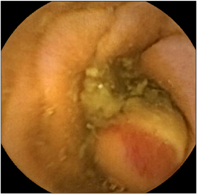
Fig. 1 Capsule endoscopic finding. It shows a suspicious huge ulcerative hyperemic mass that located in about 1 hour apart from duodenal second portion.
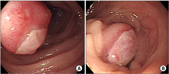
Fig. 2 Single balloon enteroscopic findings. (A) It shows a hyperemic ulcerative mass at proximal jejunum, and (B) the round central ulceration is covered with exudates.
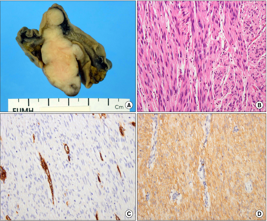
Fig. 3 Pathologic findings. (A) There is a 4.4×2.2 cm sized dumbbell shaped polypoid mass with well defined margin. (B) It is composed of spindle cells with a little mitotic activity (1 mitosis/50 HPF; H&E, ×400). (C) The staining of CD 34 is negative. (D) The staining of CD 117 (c-kit) is positive.
Reference
-
1. Cho NY, Chun HJ, Kim YS, Jeen YT, Um SH, Kim CD, et al. Chronic bleeding due to jejunal gastrointestinal stromal tumor diagnosed by capsule endoscopy. Korean J Gastrointest Endosc. 2004. 28:141–145.2. Jhu IK, Joo YE, Park GS, Park MH, Park SU, Lee NH, et al. A case of duodenal gastrointestinal stromal tumor presenting with gastrointestinal bleeding. Korean J Gastrointest Endosc. 2005. 31:121–125.3. Gourgiotis S, Kotoulas D, Aloizos S, Kolovou A, Salemis NS, Kantounakis I. Preoperative diagnosis of obscure gastrointestinal bleeding due to a GIST of the jejununm: a case report. Cases J. 2009. 2:9088.4. Kwon HJ, Cho HG, Kim MH, Ko GJ, Jeong JH, Song JS. A case of a duodenal gastrointestinal stromal tumor with a bleeding ulcer. Korean J Gastrointest Endosc. 2009. 39:42–45.5. Lee JI, Song KY, Park CH, Kim SN, Lee IS. Hemorrhagic small bowel tumor diagnosed with using capsule endoscopy and it was treated with laparoscopic surgery: report of a case. Korean J Gastrointest Endosc. 2006. 32:53–56.6. Kim HJ, Shim CS, Lee SH, Jung IS, Hong SJ, Ryu CB, et al. The usefulness of capsule endoscopy in patients with obscure gastrointestinal bleeding. Korean J Gastrointest Endosc. 2005. 31:140–146.7. Spada C, Riccioni ME, Familiari P, Marchese M, Bizzotto A, Costamagna G. Video capsule endoscopy in small-bowel tumors: a single center experience. Scand J Gastroenterol. 2008. 43:497–505.8. Rondonotti E, Pennazio M, Toth E, Menchen P, Riccioni ME, De Palma GD, et al. Small-bowel neoplasms in patients undergoing video capsule endoscopy: a multicenter European study. Endoscopy. 2008. 40:488–495.9. Bailey AA, Debinski HS, Appleyard MN, Remedios ML, Hooper JE, Walsh AJ, et al. Diagnosis and outcome of small bowel tumors found by capsule endoscopy: a three-center Australian experience. Am J Gastroenterol. 2006. 101:2237–2243.10. Postgate A, Despott E, Burling D, Gupta A, Phillips R, O'Beirne J, et al. Significant small bowel lesions detected by alternative diagnostic modalities after negative capsule endoscopy. Gastrointest Endosc. 2008. 68:1209–1214.11. Schwartz GD, Barkin JS. Small-bowel tumors detected by wireless capsule endoscopy. Dig Dis Sci. 2007. 52:1026–1030.12. Kameda N, Higuchi K, Shiba M, Machida H, Okazaki H, Yamagami H, et al. A prospective single-blind trial comparing wireless capsule endoscopy and double-balloon enteroscopy in patients with obscure gastrointestinal bleeding. J Gastroenterol. 2008. 43:434–440.13. Jang HJ, Park CH, Han SY, Byun HW, Choi MH, Kae SH, et al. Comparison of double balloon enteroscopy and capsule endoscopy for patients with suspected small bowel diseases. Korean J Gastrointest Endosc. 2007. 35:379–384.14. Chong AK, Chin BW, Meredith CG. Clinically sigificant small-bowel pathology indentified by double-balloon enteroscopy but missed by capsule endoscopy. Gastrointest Endosc. 2006. 64:445–449.15. Almeida N, Figueiredo P, Lopes S, Gouveia H, Leitão MC. Double-balloon enteroscopy and small bowel tumors: a South European single-center experience. Dig Dis Sci. 2009. 54:1520–1524.16. Lee Y, Jang JY, Ha SH, Dong SH, Kim HJ, Kim BH, et al. A case of primary jejunal mucinous adenocarcinoma diagnosed by single balloon enteroscopy. Korean J Gastrointest Endosc. 2010. 40:199–202.
- Full Text Links
-
- Actions
-
Cited
- CITED
-
- Close
- Share
-
- Similar articles
-
- A Case of Bleeding from a Jejunal Gastrointestinal Stromal Tumor Diagnosed by Double Balloon Enteroscopy
- Does Single Balloon Enteroscopy Have Similar Efficacy and Endoscopic Performance Compared with Double Balloon Enteroscopy?
- A Case of Massive Bleeding from Jejunal Stromal Tumor Diagnosed by Intraoperative Enteroscopy: A Case of Jejunal Stromal Tumor Bleeding
- A Case of Gastrointestinal Stromal Tumor of the Proximal Jejunum Diagnosed by Double Balloon Enteroscopy
- A Case of Proximal Jejunal Diverticular Bleeding Diagnosed by Double Balloon Enteroscopy and Treated by Colonoscopic Hemoclipping
