Oncocytic Sialolipoma of the Submandibular Gland
- Affiliations
-
- 1Department of Otolaryngology-Head and Neck Surgery, Kyungpook National University Hospital, Kyungpook National University School of Medicine, Daegu, Korea. entgodlikeu@gmail.com
- 2Department of Pathology, Kyungpook National University Hospital, Kyungpook National University School of Medicine, Daegu, Korea.
Abstract
- Sialolipoma, a rare tumor of the salivary gland, is a recently described variant of salivary gland lipoma. Oncocytic sialolipoma was first described by Pusiol et al. in 2009. We report the case of an oncocytic sialolipoma of the submandibular gland in a 43-year-old female. Excision of the tumor was performed with preservation of the submandibular gland. The tumor had a thin, fibrous capsule and consisted of abundant adipose tissue, an oncocytic nodule, and scattered normal glandular structures surrounded by adipose tissue. Four cases of sialolipoma of the submandibular gland, including the present case, were reviewed. All 4 tumors were developed on the right submandibular glands, with a composition of adipose tissue as high as that of sialolipoma of the parotid gland; in contrast to previous reports, three cases were in females. As newly described tumor type, care should be taken to distinguish oncocytic sialolipoma from other salivary gland neoplasms such as simple lipoma, pleomorphic adenoma, or oncocytoma.
Keyword
MeSH Terms
Figure
-

Fig. 1 (A) Ultrasonography showing that the mass (arrows) was of a heterogeneous, hypoechoic nature with ill-defined margins compared to typical submandibular gland (SMG) tissue; the mass was located throughout the superficial and deep portions of normal submandibular gland tissue (MH, mylohyoid muscle; HG, hyoglossus muscle). (B) The computed tomography image revealed a fatty mass with irregular margins and multiple soft tissue density was seen inside the mass in the right parapharyngeal space and submandbular region.
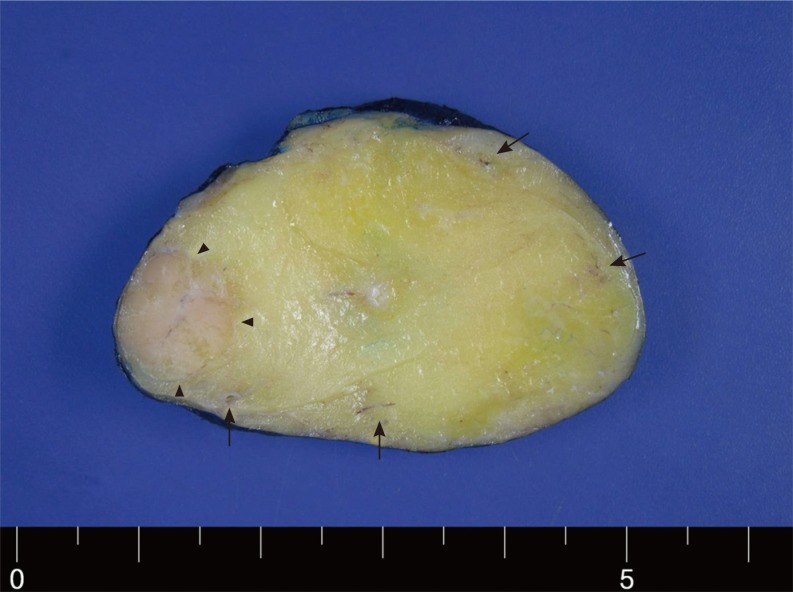
Fig. 2 Gross pathology. The tumor was well-circumscribed, soft, yellowish and had a well-demarcated light-pink colored nodular component (arrow heads) surrounded by fat tissue and ill-defined brown lesions (arrows) scattered peripherally in the tumor; histological examination identified these structures as an oncocytic nodule and glandular tissue.

Fig. 3 (A) The tumor was encapsulated by thin fibrous capsule (arrow), and the majority of the tumor consisted of adipocytes. Salivary gland tissue scattered throughout the tumor and surrounded by adipocytes. An oncocytic nodule (O) was found surrounded by abundant adipose tissue and adjacent to glandular structures (H&E, ×20). (B) The oncocytic nodule was composed of large round to polygonal cells with fine-granular eosinophilic cytoplasm and round vesicular nuclei with occasional nucleoli (H&E, ×200).
Reference
-
1. Ilie M, Hofman V, Pedeutour F, Attias R, Santini J, Hofman P. Oncocytic lipoadenoma of the parotid gland: immunohistochemical and cytogenetic analysis. Pathol Res Pract. 2010; 1. 206(1):66–72. PMID: 19346081.
Article2. Nagao T, Sugano I, Ishida Y, Asoh A, Munakata S, Yamazaki K, et al. Sialolipoma: a report of seven cases of a new variant of salivary gland lipoma. Histopathology. 2001; 1. 38(1):30–36. PMID: 11135044.
Article3. Jang YW, Kim SG, Pai H, Park JW, Lee YC, Rotaru H. Sialolipoma: case report and review of 27 cases. Oral Maxillofac Surg. 2009; 6. 13(2):109–113. PMID: 19347375.
Article4. Pusiol T, Franceschetti I, Scialpi M, Piscioli I. Oncocytic sialolipoma of the submandibular gland with sebaceous differentiation: a new pathological entity. Indian J Pathol Microbiol. 2009; Jul-Sep. 52(3):379–382. PMID: 19679967.
Article5. Ramer N, Lumerman HS, Ramer Y. Sialolipoma: report of two cases and review of the literature. Oral Surg Oral Med Oral Pathol Oral Radiol Endod. 2007; 12. 104(6):809–813. PMID: 17482843.
Article6. Yum DJ, Kang JH, Kim YJ, Kim SW. Sialolipoma of the parotid gland. Korean J Otorhinolaryngol-Head Neck Surg. 2008; 4. 51(4):371–375.7. Parente P, Longobardi G, Bigotti G. Hamartomatous sialolipoma of the submandibular gland: case report. Br J Oral Maxillofac Surg. 2008; 10. 46(7):599–600. PMID: 18374461.
Article
- Full Text Links
-
- Actions
-
Cited
- CITED
-
- Close
- Share
-
- Similar articles
-
- A Case of Oncocytic Lipoadenoma Arising in the Submandibular Gland
- A Case of Oncocytic Carcinoma Arising in the Submandibular Gland
- Sialolipoma of the Partoid Gland
- A Case of Unilateral Absence of the Submandibular Gland Secondary to Sialolithiasis
- A Case of Lymphoepithelial Carcinoma Originating in the Submandibular Gland
