Protective Effect of Metformin on Gentamicin-Induced Vestibulotoxicity in Rat Primary Cell Culture
- Affiliations
-
- 1Department of Otolaryngology-Head and Neck Surgery, Korea University College of Medicine, Seoul, Korea. logopas@korea.ac.kr
- KMID: 2278369
- DOI: http://doi.org/10.3342/ceo.2014.7.4.286
Abstract
OBJECTIVES
One of the antidiabetic drugs, metformin, have shown that it prevented oxidative stress-induced death in several cell types through a mechanism involving the opening of the permeability transition pore and cytochrome c release. Thus, it is possible that the antioxidative effect of metformin can also serve as protection against gentamicin-induced cytotoxicity related to reactive oxygen species (ROS). The aim of this study was to examine the protective effect of metformin on gentamicin-induced vestibulotoxicity in primary cell culture derived from rat utricle.
METHODS
For vestibular primary cell culture, rat utricles were dissected and incubated. Gentamicin-induced cytotoxicity was measured in both the auditory and vestibular cells. To examine the effects of metformin on gentamicin-induced cytotoxicity in the primary cell culture, the cells were pretreated with metformin at a concentration of 1 mM for 24 hours, and then exposed to 2.5 mM gentamicin for 48 hours. The intracellular ROS level was measured using a fluorescent dye, and also measured using a FACScan flow cytometer. Intracellular calcium levels in the vestibular cells were measured with calcium imaging using Fura-2 AM.
RESULTS
Vestibular cells were more sensitive to gentamicin-induced cytotoxicity than auditory hair cells. Metformin protects against gentamicin-induced cytotoxicity in vestibular cells. Metformin significantly reduced a gentamicin-induced increase in ROS, and also reduced an increase in intracellular calcium concentrations in gentamicin-induced cytotoxicity.
CONCLUSION
Metformin significantly reduced a gentamicin-induced increase in ROS, stabilized the intracellular calcium concentration, and inhibited gentamicin-induced apoptosis. Thus, Metformin showed protective effect on gentamicin-induced cytotoxicity in vestibular primary cell culture.
MeSH Terms
Figure
-

Fig. 1 Vestibular primary cell culture. (A) Rat utricles were removed for primary cell culture. Otoconia were gently removed from the utricles and the utricles were gently opened and fixed at the bottom of culture plates. Spreading vestibular cells (arrows) from the utricles were observed after 2 days of incubation. (B-D) Vestibular cells were spindle-shaped, and spreading to whole culture plate. Magnifications were ×40, ×40, ×100, and ×200, respectively.

Fig. 2 Comparison of gentamicin (GM)-induced cytotoxicity between the auditory (HEI-OC1) cell line and vestibular primary cell culture. (A) GM-induced cytotoxicity in the auditory cell line. Cell viability was 45.5% at the GM concentration of 4 mM for 48 hours. (B) GM-induced cytotoxicity in the primary cell culture. Cell viability was 57.3% at the GM concentration of 2.5 mM for 48 hours. (C) Ratio of relative viability between the auditory cell line and primary cell culture. The higher the concentration of GM was applied to cell cultures, the more cells were damaged. A trend line showed a steady ratio between the auditory cell line and primary cell culture, and vestibular cells were more sensitive to GM-induced cytotoxicity. Results from 5 separate experiments in triplicate.
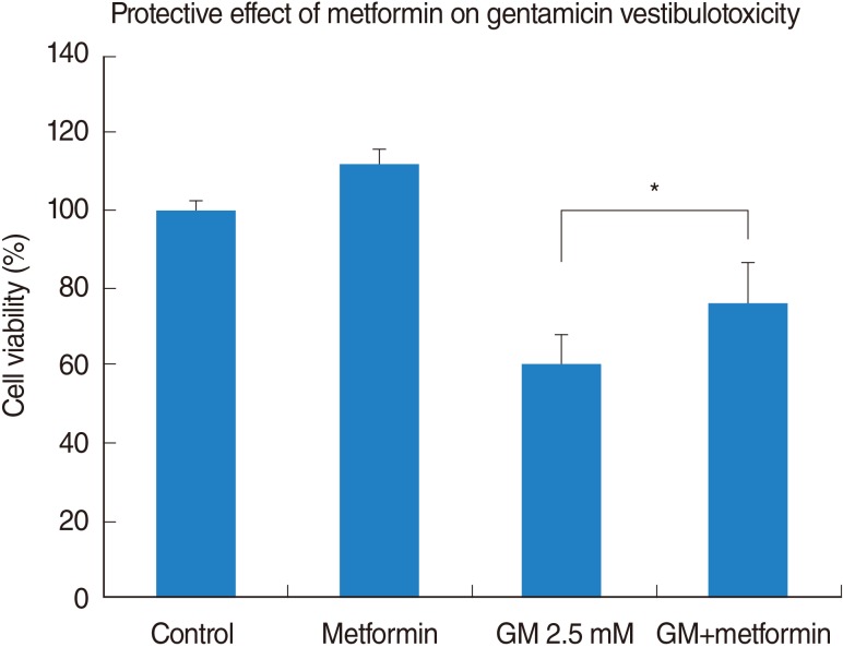
Fig. 3 Protective effect of metformin measured by the MTT assay. In the group receiving 1 mM metformin, metformin promoted slight cellular growth, showing maximal cell viability of 111.8% at the concentration of 1 mM metformin (no significant compared with control, P>0.05). Metformin provided significant protection (59.9%±3.7% for the gentamicin [GM] group vs. 75.6%±5% for the GM-plus-metformin group, mean±SE, n=5) against the toxic effect of 2.5 mM GM applied for 48 hours (*P<0.05, compared with the GM group). Results from 5 separate experiments in triplicate.

Fig. 4 Representative microscopic appearance: protective effect of metformin on gentamicin-induced cytotoxicity in vestibular primary cell culture. (A) Control group, normal vestibular primary cell culture. (B) Group receiving 1 mM metformin. Metformin promoted slight cellular growth, showing similar appearance with the control group. (C) Group receiving 2.5 mM gentamicin. Many necrotic whitish cell bodies floated on the surface of cell culture plates. The remaining cells showed condensation and shrinkage with nuclear fragmentation. (D) Gentamicin-plus-metformin group. Metformin protected against gentamicin-induced cytotoxicity. Compared with the gentamicin group, the cell size was within the normal range and necrotic bodies were fewer. However, more cells with nuclear fragmentation and necrosis were observed compared with the control group.

Fig. 5 Measurement of intracellular reactive oxygen species (ROS) production (DCFH-DA, green). (A) Control, (B) gentamicin (GM), (C) GM plus metformin, (D) vestibular cells were treated with 2.5 mM GM in the presence or absence of 1 mM metformin for 48 hours. Metformin significantly protected vestibular cells from GM-induced cytotoxicity, and ROS production in the GM-plus-metformin group was significantly lower than that in the GM and H2O2 groups (**P<0.01, ***P<0.001, respectively). However, ROS production was not significantly different between the control, metformin, an GM-plus-metformin groups (control, vestibular cells in control media; GM, cells treated with 2.5 mM GM for 48 hours; metformin, cells treated with 1 mM metformin; GM plus metformin, cells treated with 2.5 mM GM and 1 mM metformin for 48 hours; H2O2, 100 µM H2O2 was used as positive control). Results from 5 separate experiments in triplicate.
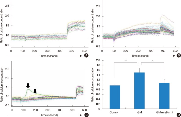
Fig. 6 Representative data of calcium imaging shows protective effects on gentamicin (GM)-induced cytotoxicity. (A) In control, no changes of calcium concentration were observed in vestibular cells. Responses to ionomycin were normal. (B) Direct application of commercial GM at concentration of 83.7 mM induced an abrupt increase in intracellular calcium concentrations in all cells tested, comparable to the level of maximal response to ionomycin. (C) In the GM-plus-metformin group, 2 cells showed an abrupt increase in calcium concentrations followed by a return to baseline levels (arrows), in contrast to other cells that had a slight and stable increase in calcium levels. (D) A before-and-after comparison shows that an increase in intracellular calcium levels was observed in the GM group compared with control (**P<0.01), but metformin reduced intracellular calcium elevation in GM-induced cytotoxicity (*P<0.05). The ratio of change in the calcium concentration was not different between the control and GM-plus-metformin groups. Results from 3 separate experiments, each experiment consisting of 20 cells.
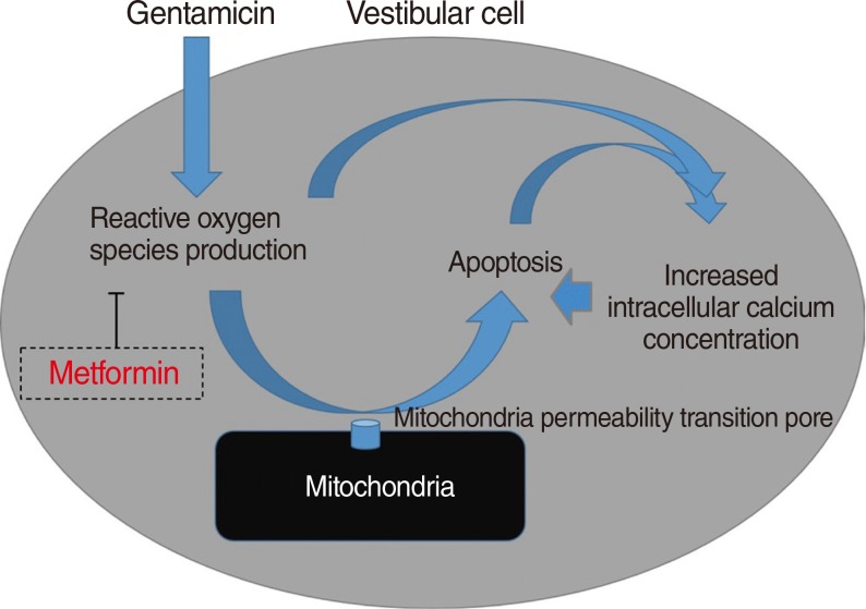
Fig. 7 Schematic mechanism on gentamicin-induced cytotoxicity. Gentamicin may enhance the formation of reactive oxygen species (ROS), which induce the opening of the mitochondrial permeability transition (MPT) pore. The MPT pore is involved in the intrinsic apoptotic pathway, and pore is opened resulting in a swelling of mitochondria and destruction of the outer mitochondrial membrane. The presence of ROS and resulting apoptosis can lead to increased intracellular calcium concentrations. Metformin significantly reduced a gentamicin-induced increase in ROS, inhibited gentamicin-induced apoptosis, and stabilized the intracellular calcium concentration.
Reference
-
1. Sajjadi H, Paparella MM. Meniere's disease. Lancet. 2008; 8. 372(9636):406–414. PMID: 18675691.
Article2. Schuknecht HF. Ablation therapy in the management of Meniere's disease. Acta Otolaryngol Suppl. 1957; 132:1–42. PMID: 13457879.3. Pullens B, van Benthem PP. Intratympanic gentamicin for Meniere's disease or syndrome. Cochrane Database Syst Rev. 2011; 3. (3):CD008234. PMID: 21412917.4. Huon LK, Fang TY, Wang PC. Outcomes of intratympanic gentamicin injection to treat Meniere's disease. Otol Neurotol. 2012; 7. 33(5):706–714. PMID: 22699980.5. Blakley BW. Update on intratympanic gentamicin for Meniere's disease. Laryngoscope. 2000; 2. 110(2):236–240. PMID: 10680922.
Article6. Minor LB. Intratympanic gentamicin for control of vertigo in Meniere's disease: vestibular signs that specify completion of therapy. Am J Otol. 1999; 3. 20(2):209–219. PMID: 10100525.7. Pender DJ. Gentamicin tympanoclysis: effects on the vestibular secretory cells. Am J Otolaryngol. 1985; Sep-Oct. 6(5):358–367. PMID: 3907388.
Article8. Beck C. Intratympanic application of gentamicin for treatment of Meniere's disease. Keio J Med. 1986; 3. 35(1):36–41. PMID: 3747323.
Article9. Tange RA, Conijn EA, van Zeijl LG, Huizing EH. Pattern of gentamicin-induced cochlear degeneration in the guinea pig: a morphological and electrophysiological study. Arch Otorhinolaryngol. 1982; 236(2):173–184. PMID: 7150082.10. Quaranta A, Scaringi A, Aloidi A, Quaranta N, Salonna I. Intratympanic therapy for Meniere's disease: effect of administration of low concentration of gentamicin. Acta Otolaryngol. 2001; 4. 121(3):387–392. PMID: 11425206.11. Dehne N, Rauen U, de Groot H, Lautermann J. Involvement of the mitochondrial permeability transition in gentamicin ototoxicity. Hear Res. 2002; 7. 169(1-2):47–55. PMID: 12121739.
Article12. Jacotot E, Costantini P, Laboureau E, Zamzami N, Susin SA, Kroemer G. Mitochondrial membrane permeabilization during the apoptotic process. Ann N Y Acad Sci. 1999; 887:18–30. PMID: 10668461.
Article13. Bernardi P, Scorrano L, Colonna R, Petronilli V, Di Lisa F. Mitochondria and cell death: mechanistic aspects and methodological issues. Eur J Biochem. 1999; 9. 264(3):687–701. PMID: 10491114.
Article14. Brustovetsky N, Brustovetsky T, Purl KJ, Capano M, Crompton M, Dubinsky JM. Increased susceptibility of striatal mitochondria to calcium-induced permeability transition. J Neurosci. 2003; 6. 23(12):4858–4867. PMID: 12832508.
Article15. Bernardi P, Krauskopf A, Basso E, Petronilli V, Blachly-Dyson E, Di Lisa F, et al. The mitochondrial permeability transition from in vitro artifact to disease target. FEBS J. 2006; 5. 273(10):2077–2099. PMID: 16649987.
Article16. Fontaine E, Eriksson O, Ichas F, Bernardi P. Regulation of the permeability transition pore in skeletal muscle mitochondria: modulation by electron flow through the respiratory chain complex I. J Biol Chem. 1998; 5. 273(20):12662–12668. PMID: 9575229.17. Hirose K, Westrum LE, Stone JS, Zirpel L, Rubel EW. Dynamic studies of ototoxicity in mature avian auditory epithelium. Ann N Y Acad Sci. 1999; 11. 884:389–409. PMID: 10842609.
Article18. Ding D, Stracher A, Salvi RJ. Leupeptin protects cochlear and vestibular hair cells from gentamicin ototoxicity. Hear Res. 2002; 2. 164(1-2):115–126. PMID: 11950531.19. El-Mir MY, Detaille D, R-Villanueva G, Delgado-Esteban M, Guigas B, Attia S, et al. Neuroprotective role of antidiabetic drug metformin against apoptotic cell death in primary cortical neurons. J Mol Neurosci. 2008; 34(1):77–87. PMID: 18040888.
Article20. Guigas B, Detaille D, Chauvin C, Batandier C, De Oliveira F, Fontaine E, et al. Metformin inhibits mitochondrial permeability transition and cell death: a pharmacological in vitro study. Biochem J. 2004; 9. 382(Pt 3):877–884. PMID: 15175014.
Article21. Kukidome D, Nishikawa T, Sonoda K, Imoto K, Fujisawa K, Yano M, et al. Activation of AMP-activated protein kinase reduces hyperglycemia-induced mitochondrial reactive oxygen species production and promotes mitochondrial biogenesis in human umbilical vein endothelial cells. Diabetes. 2006; 1. 55(1):120–127. PMID: 16380484.
Article22. Hou X, Song J, Li XN, Zhang L, Wang X, Chen L, et al. Metformin reduces intracellular reactive oxygen species levels by upregulating expression of the antioxidant thioredoxin via the AMPK-FOXO3 pathway. Biochem Biophys Res Commun. 2010; 5. 396(2):199–205. PMID: 20398632.
Article23. Algire C, Moiseeva O, Deschenes-Simard X, Amrein L, Petruccelli L, Birman E, et al. Metformin reduces endogenous reactive oxygen species and associated DNA damage. Cancer Prev Res (Phila). 012; 4. 5(4):536–543. PMID: 22262811.
Article24. Im GJ, Chang JW, Choi J, Chae SW, Ko EJ, Jung HH. Protective effect of Korean red ginseng extract on cisplatin ototoxicity in HEI-OC1 auditory cells. Phytother Res. 2010; 4. 24(4):614–621. PMID: 20020438.
Article25. Chang J, Jung HH, Yang JY, Choi J, Im GJ, Chae SW. Protective role of antidiabetic drug metformin against gentamicin induced apoptosis in auditory cell line. Hear Res. 2011; 12. 282(1-2):92–96. PMID: 21979311.26. Choi J, Im GJ, Chang J, Chae SW, Lee SH, Kwon SY, et al. Protective effects of apocynin on cisplatin-induced ototoxicity in an auditory cell line and in zebrafish. J Appl Toxicol. 2013; 2. 33(2):125–133. PMID: 22147442.
Article27. Chang J, Yang JY, Choi J, Jung HH, Im GJ. Calcium imaging in gentamicin ototoxicity: increased intracellular calcium relates to oxidative stress and late apoptosis. Int J Pediatr Otorhinolaryngol. 2011; 12. 75(12):1616–1622. PMID: 22015113.
Article28. Orrenius S, McConkey DJ, Nicotera P. Role of calcium in toxic and programmed cell death. Adv Exp Med Bio. 1991; 283:419–425. PMID: 1648868.
Article
- Full Text Links
-
- Actions
-
Cited
- CITED
-
- Close
- Share
-
- Similar articles
-
- Effect of Low Power Laser Irradiation on Gentamicin-Damaged Vestibular System in Guinea Pigs
- The Effect of Memantine on Experimentally Gentamicin Induced Vestibulotoxicity in Guinea Pigs
- Evaluation and Prevention of Gentamicin-induced Vestibulotoxicity in Rabbits Using Off-Vertical Axis Rotation
- The Protective Effect of Epigallocatechin-3-Gallate Against Gentamicin Vestibular Ototoxicity in Type I Vestibular Hair Cell of Guinea Pig
- A Promotive Effect of Low-Level Laser on Hair Cell Regeneration Following Gentamicin Induced Ototoxicity in Postnatal Organotypic Culture of Rat Utricles
