Alternative Method of Retrocrural Approach during Celiac Plexus Block Using a Bent Tip Needle
- Affiliations
-
- 1Department of Anesthesiology and Pain Medicine, Anesthesia and Pain Research Institute, Yonsei University College of Medicine, Seoul, Korea. YWLEEPAIN@yuhs.ac
- KMID: 2278270
- DOI: http://doi.org/10.3344/kjp.2015.28.2.109
Abstract
- BACKGROUND
This study sought to determine safe ranges of oblique angle, skin entry point and needle length by reviewing computed tomography (CT) scans and to evaluate the usefulness of a bent tip needle during celiac plexus block (CPB).
METHODS
CT scans of 60 CPB patients were reviewed. Image of the uppermost margin of L2 vertebral body was used to measure the minimal and maximal oblique angles and the distances from the midline to skin puncture point. The imaginary needle trajectory distance was calculated by three-dimensional measurement. When the procedure was performed by using a 10degrees bent tip needle under a 20degrees oblique X-ray fluoroscopic view, the distance (GF/G'F) from the midline to the actual puncture site was measured.
RESULTS
The imaginary safe oblique angle range was 26.4-34.2degrees and 27.7-36.0degrees on the right and left, respectively. The distance from the midline to skin puncture point was 6.1-7.6 cm on the right and 6.3-7.6 cm on the left. The needle trajectory distance at minimal angle was 9.6-11.6 cm on the right and 9.5-11.5 cm on the left. The distance of GF/G'F was 5.1-6.5 cm and 5.0-6.4 cm on the right and left, respectively. All imaginary parameters were correlated with BMI except for GF/G'F. All complications were mild and transient.
CONCLUSIONS
We identified safe values of angles and distances using a straight needle. Furthermore, using a bent tip needle under a 20degrees oblique fluoroscopic view, we could safely perform CPB with smaller parameter values.
MeSH Terms
Figure
-
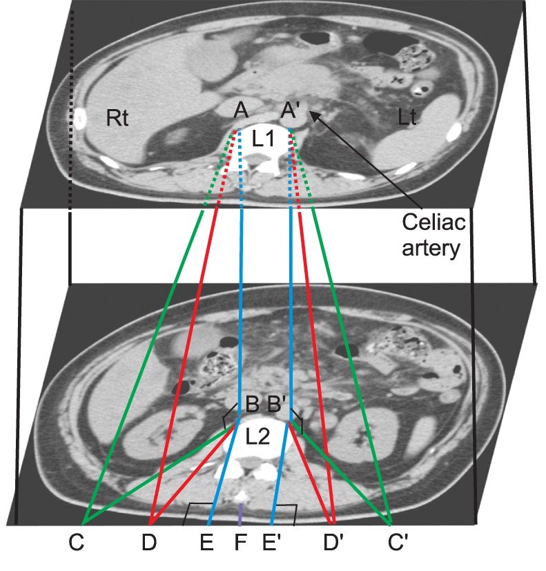
Fig. 1 Transaxial CT abdomen images of L1 vertebral body at the level of celiac artery and at the uppermost level of the L2 vertebral body. A (A'), the anterolateral part of the L1 vertebral body crossed with a vertical line of the mid portion of the pedicle; B (B'), the upper anterolateral part of the L2 vertebral body crossing the vertical line of the mid portion of the pedicle, not to penetrate the crura; C (C'), the skin surface farthest from the midline to project tangentially to B while avoiding major abdominal organs; D (D'), the nearest skin surface to project tangentially to B while avoiding interference with osteophytes or lateral bony structures of the L2 vertebral body; E (E'), the skin surface crossing the vertical line of the mid portion of the pedicle; F: midline, Rt.: right, Lt.: left.
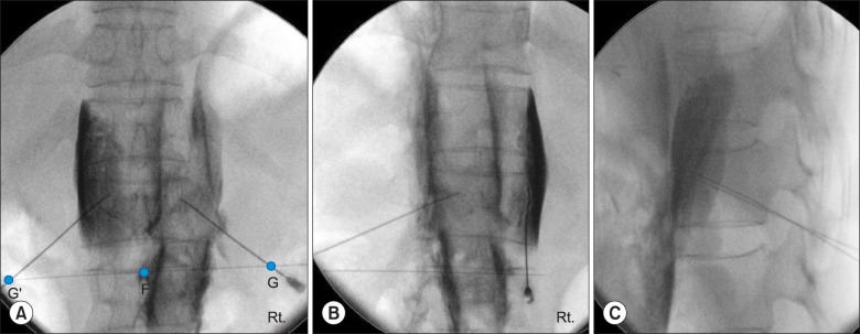
Fig. 2 X-ray fluoroscopy images ([A] Anteroposterior view, [B] Right oblique view, [C] Lateral view) of celiac plexus block by a posterior retrocrural approach that utilizes a 20° oblique angle. G (G'), the actual puncture site; F: midline, Rt.: right.
Reference
-
1. Bahn BM, Erdek MA. Celiac plexus block and neurolysis for pancreatic cancer. Curr Pain Headache Rep. 2013; 17:310. PMID: 23299904.
Article2. de Oliveira R, dos Reis MP, Prado WA. The effects of early or late neurolytic sympathetic plexus block on the management of abdominal or pelvic cancer pain. Pain. 2004; 110:400–408. PMID: 15275792.
Article3. Waldman SD, Patt RB. Splanchnic and celiac plexus nerve block. In : Waldman SD, editor. Pain management. Philadelphia (PA): Saunders-Elsevier;2006. p. 1268–1270.4. Wang PJ, Shang MY, Qian Z, Shao CW, Wang JH, Zhao XH. CT-guided percutaneous neurolytic celiac plexus block technique. Abdom Imaging. 2006; 31:710–718. PMID: 17151902.
Article5. Gress F, Schmitt C, Sherman S, Ciaccia D, Ikenberry S, Lehman G. Endoscopic ultrasound-guided celiac plexus block for managing abdominal pain associated with chronic pancreatitis: a prospective single center experience. Am J Gastroenterol. 2001; 96:409–416. PMID: 11232683.
Article6. Ward EM, Rorie DK, Nauss LA, Bahn RC. The celiac ganglia in man: normal anatomic variations. Anesth Analg. 1979; 58:461–465. PMID: 574729.7. Hur CR, Yoon DM, Oh HK, Chung MS, Chung IH. Morphological variations of the celiac plexus in Korean cadavers. J Korean Pain Soc. 1989; 2:135–144.8. De Cicco M, Matovic M, Bortolussi R, Coran F, Fantin D, Fabiani F, et al. Celiac plexus block: injectate spread and pain relief in patients with regional anatomic distortions. Anesthesiology. 2001; 94:561–565. PMID: 11379673.10. Eisenberg E, Carr DB, Chalmers TC. Neurolytic celiac plexus block for treatment of cancer pain: a meta-analysis. Anesth Analg. 1995; 80:290–295. PMID: 7818115.11. Ischia S, Ischia A, Polati E, Finco G. Three posterior percutaneous celiac plexus block techniques. A prospective, randomized study in 61 patients with pancreatic cancer pain. Anesthesiology. 1992; 76:534–540. PMID: 1550278.
Article12. Navarro-Martinez J, Montes A, Comps O, Sitges-Serra A. Retroperitoneal abscess after neurolytic celiac plexus block from the anterior approach. Reg Anesth Pain Med. 2003; 28:528–530. PMID: 14634943.
Article13. Ina H, Kitoh T, Kobayashi M, Imai S, Ofusa Y, Goto H. New technique for the neurolytic celiac plexus block: the transintervertebral disc approach. Anesthesiology. 1996; 85:212–217. PMID: 8694369.14. Singler RC. An improved technique for alcohol neurolysis of the celiac plexus. Anesthesiology. 1982; 56:137–141. PMID: 7055324.
Article15. Ischia S, Luzzani A, Ischia A, Faggion S. A new approach to the neurolytic block of the coeliac plexus: the transaortic technique. Pain. 1983; 16:333–341. PMID: 6194498.
Article16. Romanelli DF, Beckmann CF, Heiss FW. Celiac plexus block: efficacy and safety of the anterior approach. AJR Am J Roentgenol. 1993; 160:497–500. PMID: 8430543.
Article17. Lee JK, Rhee JY, Chung JG, Rhee CS. CT - guided celiac plexus block using anterior approach. J Korean Pain Soc. 1999; 12:87–94.18. Rana MV, Candido KD, Raja O, Knezevic NN. Celiac plexus block in the management of chronic abdominal pain. Curr Pain Headache Rep. 2014; 18:394. PMID: 24414338.
Article19. Oh HK. Celiac plexus block: a review. J Korean Pain Soc. 1990; 3:1–3.20. Ochiai Y, Ishii S, Takahashi S, Mizobuchi S, Fukushima T, Katayama H, et al. Appropriate puncture site for CT monitored celiac plexus block determined from CT films. Masui. 1992; 41:1961–1965. PMID: 1479665.21. Weber JG, Brown DL, Stephens DH, Wong GY. Celiac plexus block. Retrocrural computed tomographic anatomy in patients with and without pancreatic cancer. Reg Anesth. 1996; 21:407–413. PMID: 8895999.
- Full Text Links
-
- Actions
-
Cited
- CITED
-
- Close
- Share
-
- Similar articles
-
- A Modification of Transaortic Celiac Plexus Neurolysis; One Needle, Periaortic Block: A case report
- The Spread of Contrast Media in Celiac Plexus Block
- Cases Report of Classic Celiac Plexus Block and Transaortic Celiac Plexus Block
- Splanchnic Nerve Block at T12 Level
- Failed Celiac Plexus Block Via the Anterior Approach under CT Guidance
