Korean J Obstet Gynecol.
2011 Aug;54(8):464-467. 10.5468/KJOG.2011.54.8.464.
An orbital hemangioendothelioma of fetus detected by prenatal ultrasonography
- Affiliations
-
- 1Department of Obstetrics and Gynecology, Keimyung University School of Medicine, Daegu, Korea. jcpark@dsmc.or.kr
- KMID: 2274068
- DOI: http://doi.org/10.5468/KJOG.2011.54.8.464
Abstract
- Most orbital tumors of infants include retinoblastoma, dermoid cyst (teratoma), optic nerve glioma and nevus, and hemangioendothelioma is found in rare cases. Hemangioendothelioma, the tumor of intermediate malignancy between angiosarcoma and hemangioma, is commonly recognized in soft tissue of extremities, skin, lung and liver with symptoms of ulceration and hepatomegaly in neonates and infants. However it seldom localizes in the orbit. Although there have been two case reports of orbital hemangioendothelioma in neonate and adult in Korea, there was no case report of prenatally diagnosed orbital hemangioendothelioma in fetus. We found hemangioma-like orbital tumor in a fetus at 36 weeks of gestation by prenatal ultrasonography and confirmed hemangioendothelioma by microscopic examination after birth. This is the first case of orbital hemangioendothelioma in fetus.
MeSH Terms
Figure
-

Fig. 1 At 36 weeks, ultrasonography reveals 6.5 × 4.8 cm sized mass on left frontal area of fetal head.

Fig. 2 3D-ultrasonography shows heterogenous mass on left orbital area of a 36 weeks old fetus.

Fig. 3 Fetal magnetic resonance image shows a relatively well-definded, huge, heterog-enous, solid mass involving the left orbit, infratemporal fossa, epidural space of left frontal and temporal lobe of a fetus, measuring 82×62×75 mm (arrow). The mass is highly vascular, internal hemorrhagic and tiny calcified foci. Several large draining vessels are noted.
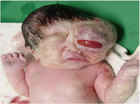
Fig. 4 After birth we defined huge orbital mass occupying left orbit and left frontotemporal area of a newborn infant.
Reference
-
1. Gull I, Wolman I, Har-Toov J, Amster R, Schreiber L, Lessing JB, et al. Antenatal sonographic diagnosis of epignathus at 15 weeks of pregnancy. Ultrasound Obstet Gynecol. 1999. 13:271–273.2. Robson CD, Barnewolt CE. MR imaging of fetal head and neck anomalies. Neuroimaging Clin N Am. 2004. 14:273–291.3. Roh KK, Lee JH, Youn DH. Clinical analysis in tumors of the eye and its adnexa. Korean J Ophthalmol. 1988. 2:27–31.4. Salim A, Wiknjosastro GH, Danukusumo D, Barnas B, Zalud I. Fetal retinoblastoma. J Ultrasound Med. 1998. 17:717–720.5. Herman TE, Vachharajani A, Siegel MJ. Massive congenital orbital teratoma. J Perinatol. 2009. 29:396–397.6. Monrigal E, Gallot D, James I, Hameury F, Vanlieferinghen P, Guibaud L. Venous malformation of the soft tissue associated with blue rubber bleb nevus syndrome: prenatal imaging and impact on postnatal management. Ultrasound Obstet Gynecol. 2009. 34:730–732.7. Sueters M, Peek AM, Ball LM, Hogendoorn PC, Scherjon SA, de Keizer RJ, et al. Prenatal detection of orbital rhabdomyosarcoma. Arch Ophthalmol. 2005. 123:276–279.8. Gupta NP, Kolla SB, Panda S, Sharma MC. Epitheloid hemangioendothelioma of urinary bladder. Indian J Urol. 2008. 24:253–255.9. Kim JS, Moon SK, Yoon HS, Lee TS. A case of infantile hemangioendothelioma of the liver treated with hepatic embolization and lobectomy. Korean J Pediatr. 2005. 48:660–664.10. Dreyfus M, Baldauf JJ, Dadoun K, Becmeur F, Berrut F, Ritter J. Prenatal diagnosis of hepatic hemangioma. Fetal Diagn Ther. 1996. 11:57–60.11. Duncan KR. The development of magnetic resonance imaging in obstetrics. Br J Hosp Med. 1996. 55:178–181.12. Roos JE, Pfiffner R, Stallmach T, Stuckmann G, Marincek B, Willi U. Infantile hemangioendothelioma. Radiographics. 2003. 23:1649–1655.13. Bergström K, Enoksson P, Gamstorp I, Naeser P. Haemangioendothelioma of the orbit. Ophthalmologica. 1978. 177:115–120.
- Full Text Links
-
- Actions
-
Cited
- CITED
-
- Close
- Share
-
- Similar articles
-
- 2 Cases of Congenital Jejunal Atresia Diagnosed by Prenatal Ultrasonography
- A Case of Fetus in Fetu: Ultrasonic prenatal diagnosis
- A Case of Cystic Lymphangioma of the Scrotum and Retroperitoneum was Detected in Fetus
- Imaging Diagnosis of Fetal Abdominal Solid Tumor
- A Case of Hemangioendothelioma Diagnosed by Antenatal Ultrasonography