Korean J Obstet Gynecol.
2011 Jul;54(7):386-389. 10.5468/KJOG.2011.54.7.386.
A case of terminal deletion of chromosome 10p
- Affiliations
-
- 1Department of Obstetrics and Gynecology, Gyeongsang National University School of Medicine, Jinju, Korea. wypaik@gnu.ac.kr
- 2Gyeongsang Institute of Health Sciences, Gyeongsang National University, Jinju, Korea.
- KMID: 2274055
- DOI: http://doi.org/10.5468/KJOG.2011.54.7.386
Abstract
- Chromosome 10p deletion (partial monosomy 10p) is rare chromosomal disorder. It was first reported in 1970. Since then, as far as we know, about 45 patients have been described. The main feature of this syndrome are craniofacial dysmorphism, congenital heart disease, vesicoureteral abnormalities, and developmental delay. We have experienced a prenatal case of Chromosome 10p terminal deletion by doing cytogenetic study due to high Down syndrome risk on quadruple test and cleft lip on prenatal ultrasonography. Conventional cytogenetic result from cord blood was 46,XY,del(10)(p13), molecular cytogenetic techniques using bacterial artificial chromosome array comparative genomic hybridization and fluorescence in situ hybridization analysis result was 46,XY,del(10)(p14)(NEBL-). To our knowledge, this karyotype may be the first report in Korea. We present this case with brief review of literature.
Keyword
MeSH Terms
-
Chromosome Deletion
Chromosome Disorders
Chromosomes, Artificial, Bacterial
Chromosomes, Human, Pair 10
Cleft Lip
Comparative Genomic Hybridization
Cytogenetic Analysis
Cytogenetics
Down Syndrome
Fetal Blood
Fluorescence
Heart Diseases
Humans
In Situ Hybridization
Karyotype
Korea
Monosomy
Ultrasonography, Prenatal
Chromosome Deletion
Chromosome Disorders
Chromosomes, Human, Pair 10
Figure
-
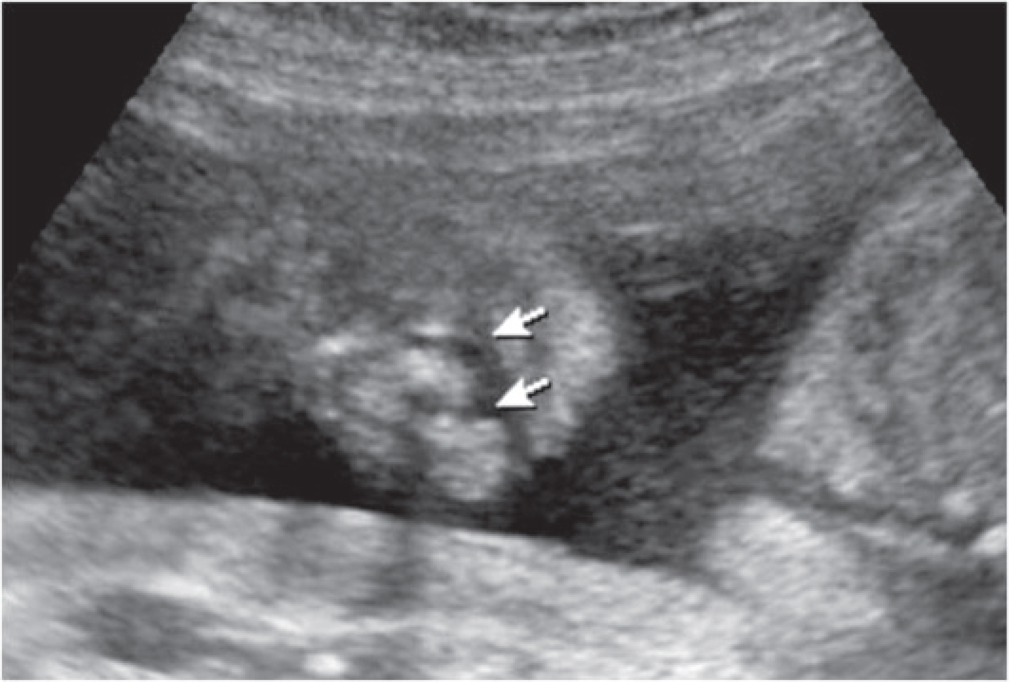
Fig. 1. Transabdominal sonography shows fetal cleft lip (arrows).
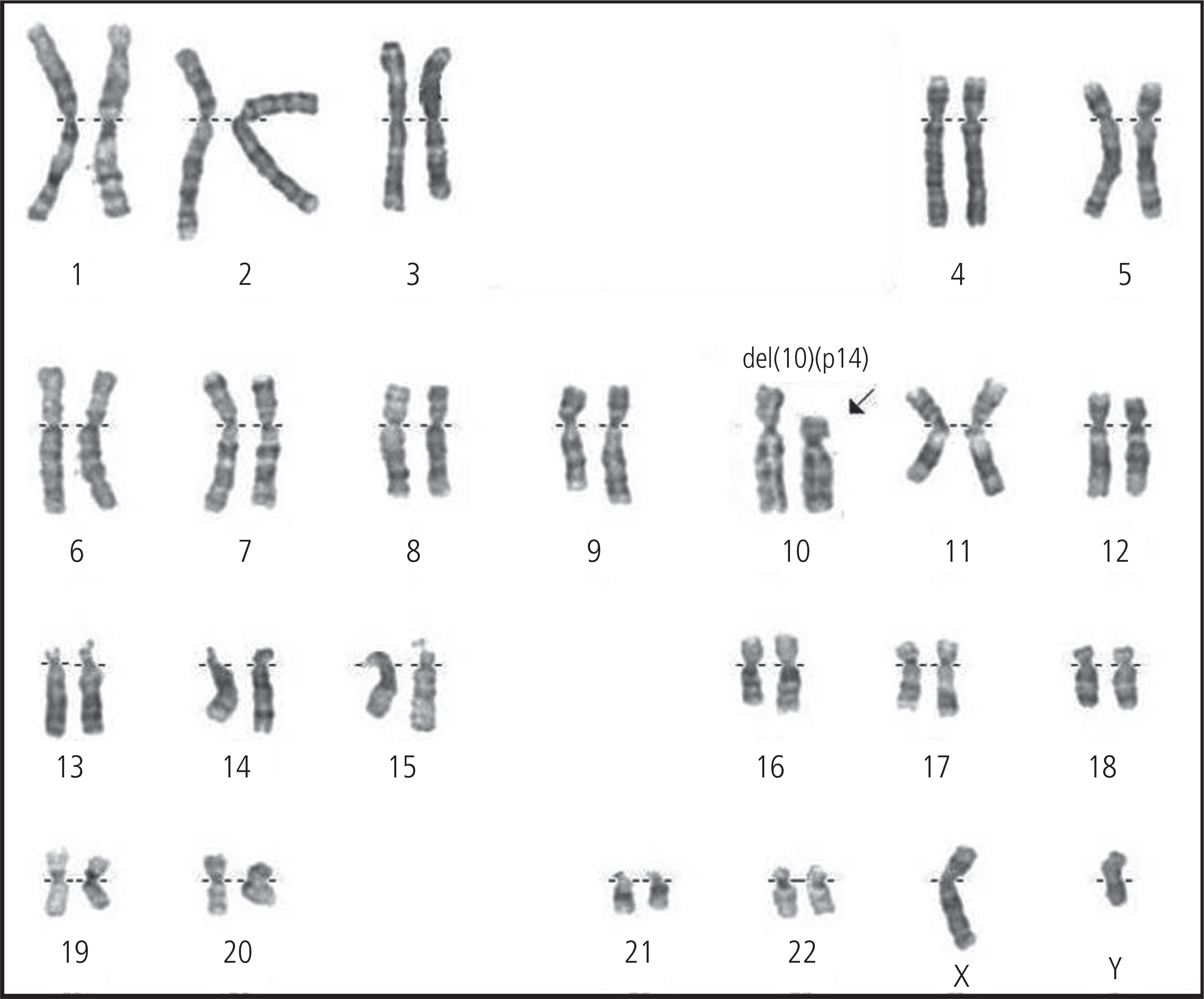
Fig. 2. Karyotype of fetus from cord blood showing 46,XY,del(10)(p13) (arrow).

Fig. 3. Bacterial artificial chromosome array comparative genomic hybridization shows deficit of DNA copy on chromosome 10 (arrow).
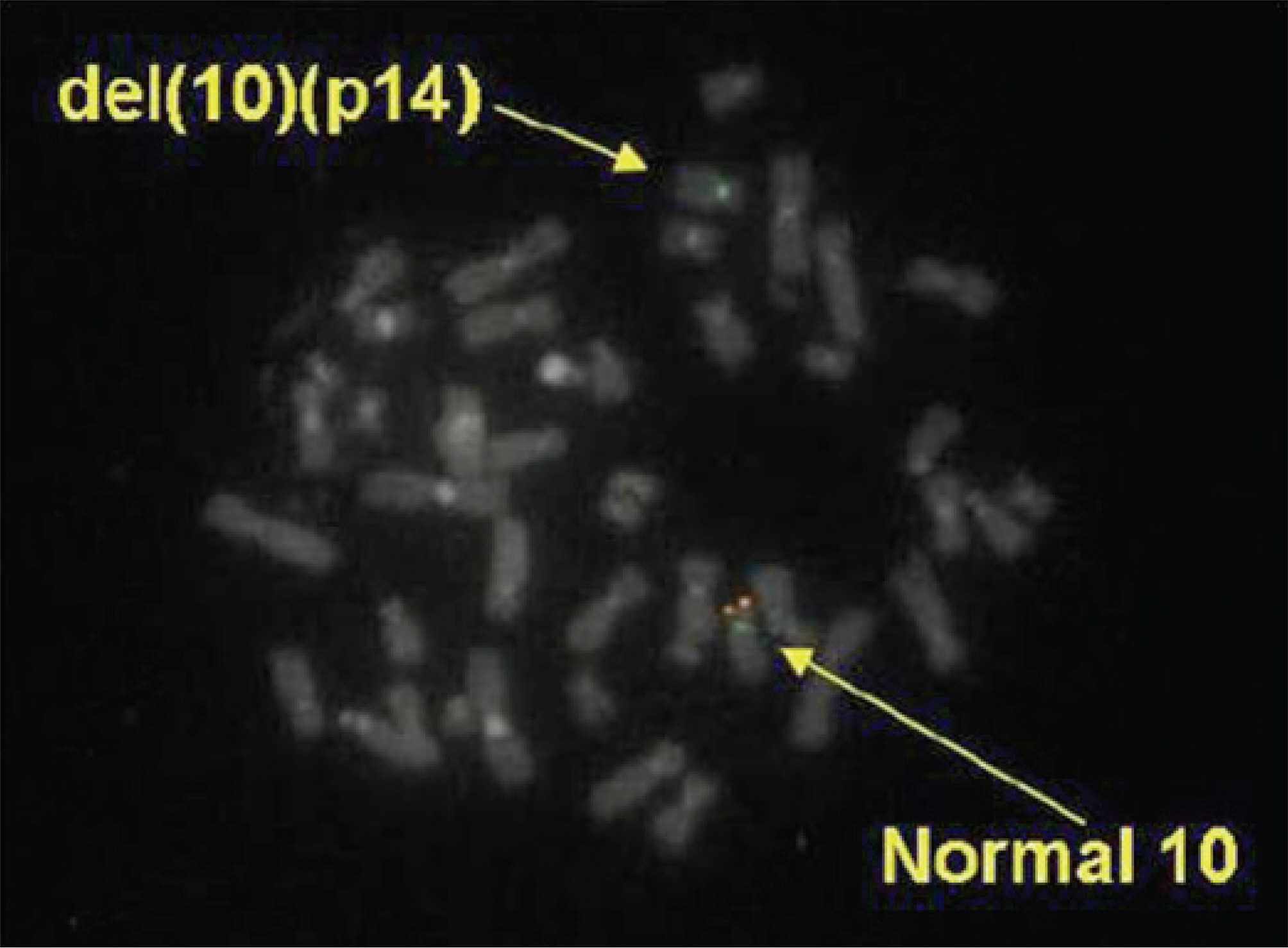
Fig. 4. Fluorescence in situ hybridization analysis. Fluorescent signal (green signal, orange signal) is detected in the distal region of the normal chromosome 10 (long arrow) and signal defect (orange signals) is showed in deleted chromosome 10 (arrow). Chromosomes were counterstained with FITC/DAPI (×1000).
Reference
-
1. Elliott D, Thomas GH, Condron CJ, Khuri N, Richardson F. C-group chromosome abnormality (? 10p-). Occurrence in a child with multiple malformations. Am J Dis Child. 1970; 119:72–3.2. Klep-de Pater JM, Bijlsma JB, Alkema FM. Partial monosomy 10p syndrome. Eur J Pediatr. 1981; 137:243–6.3. Schuffenhauer S, Seidel H, Oechsler H, Belohradsky B, Bernsau U, Murken J, et al. DiGeorge syndrome and partial monosomy 10p: case report and review. Ann Genet. 1995; 38:162–7.4. Lichtner P, König R, Hasegawa T, Van Esch H, Meitinger T, Schuffenhauer S. An HDR (hypoparathyroidism, deafness, renal dysplasia) syndrome locus maps distal to the DiGeorge syndrome region on 10p13/14. J Med Genet. 2000; 37:33–7.
Article5. Shokeir MH, Ray M, Hamerton JL, Bauder F, O'Brien H. Deletion of the short arm of chromosome No. 10. J Med Genet. 1975; 12:99–103.
Article6. Lindstrand A, Malmgren H, Verri A, Benetti E, Eriksson M, Nordgren A, et al. Molecular and clinical characterization of patients with overlapping 10p deletions. Am J Med Genet A. 2010; 152A:1233–43.
Article7. Slinde S, Hansteen IL. Two chromosomal syndromes in the same family: monosomy and trisomy for part of the short arm of chromosome 10. Eur J Pediatr. 1982; 139:153–7.
Article8. Van Esch H, Groenen P, Nesbit MA, Schuffenhauer S, Lichtner P, Vanderlinden G, et al. GATA3 haplo-insufficiency causes human HDR syndrome. Nature. 2000; 406:419–22.
Article9. Yang YH, Kang JY, Yang ES, Jang SY, Cho JS, Park YW, et al. Clinical usefulness of fluorescence in situ hybridization (FISH) in the diagnosis of genetic disease. Korean J Obstet Gynecol. 2002; 45:1016–25.
- Full Text Links
-
- Actions
-
Cited
- CITED
-
- Close
- Share
-
- Similar articles
-
- Terminal Deletion of the Chromosome 4q with Hemivertebra: Case Report
- A newborn with developmental delay diagnosed with 4q35 deletion and 10p duplication
- 6p23 Deletion Syndrome: Report of a Case in a Preterm Baby
- A Case of Trisomy 10p with Vertebral Anomaly and Hypospadias
- Partial Monosomy 21 Associated with Unbalanced t(10p; 21q)
