Congenital lymphangiomatosis of the right lower limb
- Affiliations
-
- 1Department of Obstetrics and Gynecology, The Catholic University of Korea School of Medicine, Seoul, Korea. jcshin@catholic.ac.kr
- KMID: 2273946
- DOI: http://doi.org/10.5468/kjog.2010.53.7.647
Abstract
- Lymphangiomatosis is a condition of lymphatic tissue malformation with multiple or diffuse involvement of soft tissues, visceral organs. Congenital abnormalities of the lymphatic system are very rare, and reports of congenital lymphangiomatosis are even fewer. We experienced a case of congenital lymphangiomatosis detected as edema of the right limb by prenatal ultrasonography and then diagnosed by magnetic resonance imaging. We describe this case with a brief review of the literature.
MeSH Terms
Figure
-

Fig. 1 Two dimensional and three dimensional ultrasonography images at 31+5 weeks of gestation showed fetal low limbs; right thigh (A), left and right thigh (B), left leg and foot (C) and right leg and foot (D). Right lower limb was edematous for left leg (D).

Fig. 2 Fetal MRI imaged showed edema of right lower limb; leg (A), thigh (B). Skin and subcutaneous tissue of right lower limb (a white arrow) was thicker for left lower limb (a white arrow head). A scan performed with techniques to study the accompanying abnormalities. There was no particular abnormality.
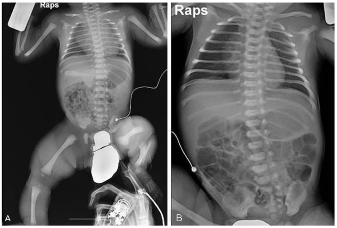
Fig. 3 Chest X-ray images of the neonatal boy showed no abnormal finding of the other side.

Fig. 4 Magnetic resonance imaging of newborn boy showed enlarged right lower limb. Diffuse edematous swelling of skin and subcutaneous soft tissue was showed with high signal on T2 weighted image (A) and T1 weighted image (B). After infusion of contrast media, thin reticular enhancements are seen in the subcutaneous fat layer (C). Photograph of the newborn boy showing enlargement and discoloration of the right lower limb (D).
Reference
-
1. Bickel WH, Brodere AC. Primary lymphangioma of the ilium; report of a case. J Bone Joint Surg Am. 1947. 29:517–522.2. Hayes JT, Brody GL. Cystic lymphangiectasis of bone: a case report. J Bone Joint Surg Am. 1961. 43:107–117.3. Laverdiere C, David M, Dubois J, Russo P, Hershon L, Lapierre JG. Improvement of disseminated lymphangiomatosis with recombinant interferon therapy. Pediatr Pulmonol. 2000. 29:321–324.4. Margraf LR. Thoracic lymphangiomatosis. Pediatr Pathol Lab Med. 1996. 16:155–160.5. Bae JH, Ahn HY, Lee JH, Kwon I, Moon HB, Kim SJ, et al. A case of prenatally diagnosed fetal retroperitoneal cystic lymphangioma. Korean J Obstet Gynecol. 2003. 46:851–855.6. Ro JY, Jung JU, Min JY, Lee HE, Jung BH, Joo IS, et al. A case of cystic lymphangioma of the scrotum and retroperitoneum was detected in fetus. Korean J Obstet Gynecol. 2004. 47:577–580.7. Kim TE, Lee SP, Park JM, Whang BC, Kim SY. Prenatal ultrasonographic diagnosis of thoracoabdominal cavernous lymphangioma: a case report. Korean J Obstet Gynecol. 2009. 52:867–871.8. Haeusler MC, Hofmann HM, Hoenigl W, Karpf EF, Rosenkranz W. Congenital generalized cystic lymphangiomatosis diagnosed by prenatal ultrasound. Prenat Diagn. 1990. 10:617–621.9. Carlson KC, Parnassus WN, Klatt EC. Thoracic lymphangiomatosis. Arch Pathol Lab Med. 1987. 111:475–477.10. Iwabuchi A, Otaka M, Okuyama A, Jin M, Otani S, Itoh S, et al. Disseminated intra-abdominal cystic lymphangiomatosis with severe intestinal bleeding. A case report. J Clin Gastroenterol. 1997. 25:383–386.11. Meredith WT, Levine E, Ahlstrom NG, Grantham JJ. Exacerbation of familial renal lymphangiomatosis during pregnancy. AJR Am J Roentgenol. 1988. 151:965–966.12. Dutheil P, Leraillez J, Guillemette J, Wallach D. Generalized lymphangiomatosis with chylothorax and skin lymphangiomas in a neonate. Pediatr Dermatol. 1998. 15:296–298.13. Shah AR, Dinwiddie R, Woolf D, Ramani R, Higgins JN, Matthew DJ. Generalized lymphangiomatosis and chylothorax in the pediatric age group. Pediatr Pulmonol. 1992. 14:126–130.14. Marymont JV, Knight PJ. Splenic lymphangiomatosis: a rare cause of splenomegaly. J Pediatr Surg. 1987. 22:461–462.15. Tran D, Fallat ME, Buchino JJ. Lymphangiomatosis: a case report. South Med J. 2005. 98:669–671.16. Gomez CS, Calonje E, Ferrar DW, Browse NL, Fletcher CD. Lymphangiomatosis of the limbs. Clinicopathologic analysis of a series with a good prognosis. Am J Surg Pathol. 1995. 19:125–133.17. Imiela A, Salle-Staumont D, Breviere GM, Catteau B, Martinot-Duquennoy V, Piette F. Congenital elephantiasis-like lymphangiomatosis of a lower limb. Acta Derm Venereol. 2003. 83:40–43.
