Term angular pregnancy with placenta accreta
- Affiliations
-
- 1Department of Obstetrics and Gynecology, Soonchunhyang University Bucheon Hospital, Soonchunhyang University College of Medicine, Bucheon, Korea. hhl22@chol.com
- 2Department of Radiology, Soonchunhyang University Bucheon Hospital, Soonchunhyang University College of Medicine, Bucheon, Korea.
- KMID: 2273892
- DOI: http://doi.org/10.5468/kjog.2010.53.6.520
Abstract
- Angular pregnancy is rare, in which the embryo in the lateral angle of uterine cavity and located medial to the utero-tubal junction. Angular pregnancy is differentiated from interstitial pregnancy. There is no report about term angular pregnancy in Republic of Korea, a few reports in other countries. Angular pregnancy has different clinical characteristics according to the trimester. We diagnosed angular pregnancy by ultrasonography and computed tomography (CT). The CT is a useful diagnostic method. We report a case of term angular pregnancy with placenta accreta and review the diagnostic process and complications.
Keyword
Figure
-
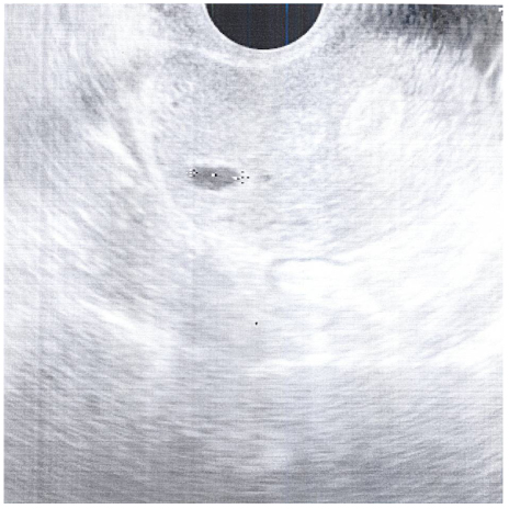
Figure 1 Ultrasonographic finding at amenorrhea 7 weeks showed gestational sac located in right upper angular area.
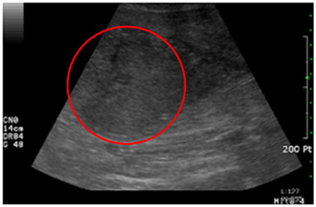
Figure 2 Ultrasonographic finding after delivery is retained placenta and placenta accreta with suspicious corneal pregnancy.
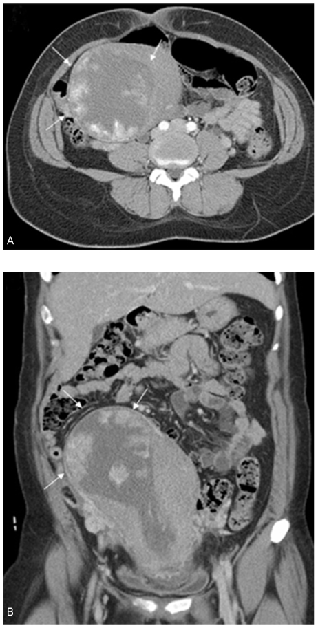
Figure 3 (A) and (B) computed tomography scan showed lobulating enhancing mass-like lesion in right upper aspect of endometrial cavity (arrows). Overlying myometrium is less than 3 mm. Retained placenta is suggested on right cornual area. Remaining portion of uterus revealed mild enlargement with normal thickness of myometrium.
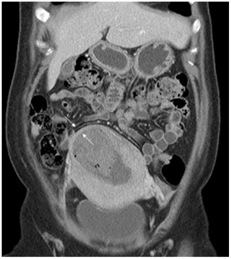
Figure 4 On the follow-up computed tomography scan after placental removal, myometrium on right cornual area is about 3.4 mm (arrows), indicating previous angular pregnancy. Hematoma is still noted within distended endometrial cavity.
Reference
-
1. Jansen RP, Elliott PM. Angular intrauterine pregnancy. Obstet Gynecol. 1981. 58:167–175.2. Kelly HA. Operative Gynecology. 1896. Baltimore: Johns Hopkins Bulletin.3. Kim HG, Park BS, Kang YP, Lee SB. A case of ruptured angular pregnancy at 26th gestational week. Korean J Obstet Gynecol. 1995. 38:2163–2167.4. Breen JL. A 21 year survey of 654 ectopic pregnancies. Am J Obstet Gynecol. 1970. 106:1004–1019.5. Divry V, Hadj S, Bordes A, Genod A, Salle B. Case of progressive intrauterine twin pregnancy after surgical treatment of cornual pregnancy. Fertil Steril. 2007. 87:190.e1. 190.e3.6. Deckers EA, Stamm CA, Naake VL, Dunn TS, McFee JG. Hysterotomy for retained placenta in a term angular pregnancy. A case report. J Reprod Med. 2000. 45:153–155.7. Tarim E, Ulusan S, Kilicdag E, Yildirim T, Bagis T, Kuscu E. Angular pregnancy. J Obstet Gynaecol Res. 2004. 30:377–379.8. Lancet M, Bin-Nun I, Kessler I. Angular and interstitial pregnancy. Int Surg. 1977. 62:10709.9. Stiller RJ, de Regt RH. Prenatal diagnosis of angular pregnancy. J Clin Ultrasound. 1991. 19:374–376.10. Filhastre M, Dechaud H, Lesnik A, Taourel P. Interstitial pregnancy: role of MRI. Eur Radiol. 2005. 15:93–95.11. Coady DJ, Snyder JR, Goldstein SR, Subramanyan BR. Ultrasound diagnosis of interstitial pregnancy. N Y State J Med. 1985. 85:655–656.12. Barron LR, Rona G, Tucker E. Ruptured angular (cornual) pregnancy. Can Med Assoc J. 1966. 95:162–163.13. Triolo O, Mancuso A, De Vivo A, Falcone S. Term angular pregnancy with placenta accreta. A case report. Clin Exp Obstet Gynecol. 2004. 31:147–148.
- Full Text Links
-
- Actions
-
Cited
- CITED
-
- Close
- Share
-
- Similar articles
-
- Angular pregnancy complicated with preterm labor at 25 gestational weeks of pregnancy
- Retained placenta accreta after a first-trimester abortion manifesting as an uterine mass
- A Case of Placenta Percreta Involving the Urinary Bladder
- A Case of placenta increta which was found about 50days after induced abortion at 1st Trimester
- The Efficacy of Color Doppler Ultrasound in Diagnosis and Management of Placenta Previa with Accreta
