Changes in longitudinal craniofacial growth in subjects with normal occlusions using the Ricketts analysis
- Affiliations
-
- 1Department of Orthodontics, School of Dentistry, Kyungpook National University, Daegu, Korea. owkwon@knu.ac.kr
- KMID: 2273430
- DOI: http://doi.org/10.4041/kjod.2014.44.2.77
Abstract
OBJECTIVE
This study was designed to define the Korean norm values for the Ricketts analysis.
METHODS
In this longitudinal study, lateral cephalograms of 31 subjects with normal occlusion were taken biennially from ages 9-19 years. Cephalometric measurements were performed. Parameters for which the 10-year change did not exceed one standard deviation were defined as unchanged. The means and standard deviations for the measured parameters were determined for each age group.
RESULTS
No significant changes in growth were observed in the molar relationship, incisor overjet, incisor overbite, mandibular incisor extrusion, interincisor angle, lower incisor tip (B1) to A point-Pogonion (A-PO) plane, upper incisor tip (A1) to A-PO plane, B1 inclination to A-PO, A1 inclination to A-PO, B1 inclination to Frankfurt plane (FH), convexity, lower facial height, facial axis, maxillary depth, maxillary height, palatal plane to FH, cranial deflection, ramus Xi position, or porion location. Continual changes over the 10 years of growth were observed in the maxillary first molar distal position to pterygoid true vertical plane, facial depth, mandibular plane to FH, anterior cranial length, mandibular arc, and corpus length.
CONCLUSIONS
Clinicians can apply the Korean norms at age 9 as determined in this study when using the Ricketts analysis. The patient's age at the beginning of treatment and their sex should be taken into consideration when drawing visual treatment objectives.
MeSH Terms
Figure
-
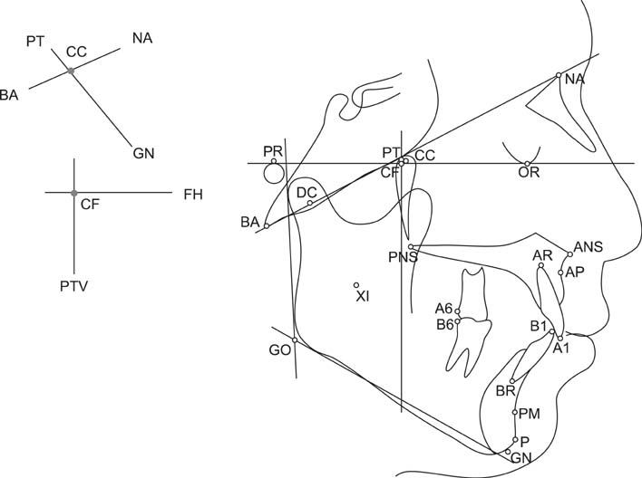
Figure 1 Anatomic and cephalometric landmarks. NA, Nasion; OR, orbitale; PR, porion; BA, basion; PT, pterygoid point; DC, condyle center point; CC, center of cranium point; CF, center of face point; ANS, anterior nasal spine; PNS, posterior nasal spine; AP, A point; XI, Xi point; PM, protuberance menti; P, pogonion; GN, gnathion; GO, gonion; A1, upper incisor tip; AR, upper incisor root tip; A6, upper first molar distal; B1, lower incisor tip; BR, lower incisor root tip; B6, lower first molar distal. FH, Frankfurt.

Figure 2 Measurements of dental relationships. A, Molar relationship; B, incisor overjet; C, incisor overbite; D, mandibular incisor extrusion; E, interincisor angle. A1, Maxillary incisor; B1, mandibular incisor; A6, maxillary first molar; B6, mandibular first molar.
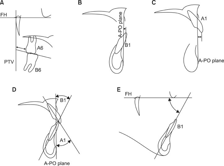
Figure 3 Measurements of dental to skeletal relationship. A, Distance of A6 to PTV, parallel to the occlusal plane; B, perpendicular distance of B1 to A-PO plane; C, perpendicular distance of A1 to A-PO plane; D, B1 inclination to A-PO and A1 inclination to A-PO; E, B1 inclination to FH. PTV, Pterygoid true vertical plane; A-PO plane, A point-Pogonion plane. See Figure 1 for the definitions of all the landmarks and measurements.

Figure 4 Measurements of skeletal relationships. A, Convexity, which indicates a perpendicular distance of A point to the facial plane; B, lower facial height, which indicates an angle between ANS-XI and XI-PM. Facial plane indicates a Nasion-Pogonion line. See Figure 1 for the definitions of all the landmarks and measurements.

Figure 5 Measurements of the jaw to cranium relationship. A, Facial depth; B, facial axis; C, maxillary depth; D, maxillary height; E, palatal plane to FH. See Figure 1 for the definitions of all the landmarks and measurements.
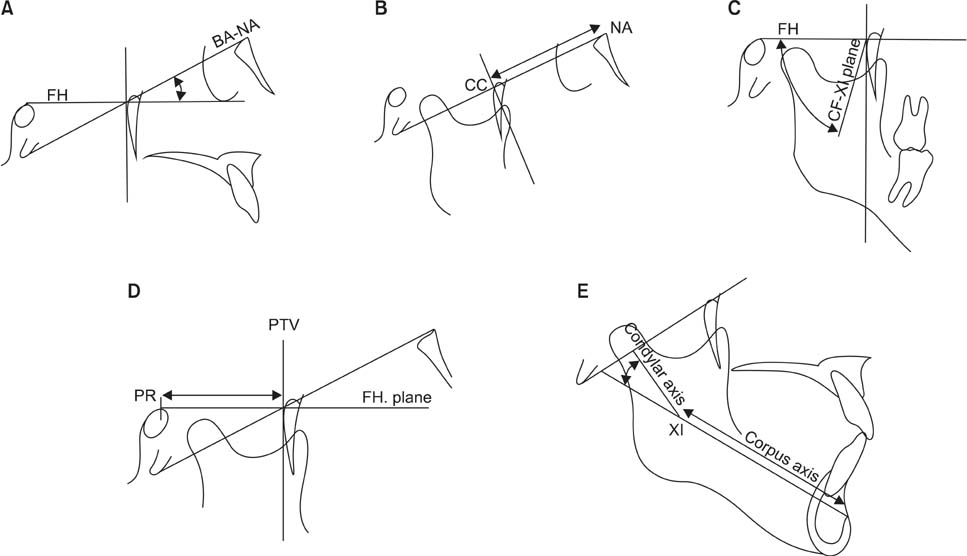
Figure 6 Measurements of internal structures. A, Cranial deflection; B, anterior cranial length; C, ramus Xi position; D, porion location; E, mandibular arc and corpus length. See Figure 1 for the definitions of all the landmarks and measurements.
Reference
-
1. Broadbent BH. A new X-ray technique and its application to orthodontia. Angle Orthod. 1931; 1:45–66.2. Downs WB. Variations in facial relationships; their significance in treatment and prognosis. Am J Orthod. 1948; 34:812–840.
Article3. Steiner CC. Cephalometrics for you and me. Am J Orthod. 1953; 39:729–755.
Article4. Sassouni V. A roentgenographic cephalometric analysis of cephalo-facio-dental relationships. Am J Orthod. 1955; 41:735–764.
Article5. Ricketts RM. A foundation for cephalometric communication. Am J Orthod. 1960; 46:330–357.
Article6. Tweed CH. Was the development of the diagnostic facial triangle as an accurate analysis based on fact or fancy? Am J Orthod. 1962; 48:823–840.
Article7. Jarabak JR. Technique and treatment with light wire edgewise appliance. 2nd ed. St. Louis: C.V. Mosby;1972. p. 128–166.8. Downs WB. Analysis of the dentofacial profile. Angle Orthod. 1956; 26:191–212.9. Sassouni V. The face in five dimensions. Philadelphia: Growth Center Publication;1960.10. Ricketts RM. A four-step method to distinguish orthodontic changes from natural growth. J Clin Orthod. 1975; 9:208–215. 218–228.11. Hamm SM, Sohn BH. Roentgenocephalometric study of craniofacial growth by Ricketts analysis on teen-ager with normal occlusion in Korean. Korean J Orthod. 1985; 15:313–326.12. Park TS. A longitudinal cephalometric study of craniofacial growth of Korean children. Korean J Orthod. 1984; 14:217–231.13. Kim YJ, Park KD, Kwon OW. A longtudinal cephalometric study of the craniofacial growth changes of adolescence with normal occlusion. Korean J Orthod. 1995; 25:287–297.14. Houston WJ. The analysis of errors in orthodontic measurements. Am J Orthod. 1983; 83:382–390.
Article15. Ricketts RM. Orthodontic diagnosis and planning. Philadelphia: Saunders;1982.16. Ricketts RM, Bench RW, Gugino CF, Hilgers JJ, Schulhof RJ. Bioprogressive therapy. Denver: Rocky Mountain/Orthodontics;1979.17. Ricketts RM. Cephalometric analysis and synthesis. Angle Orthod. 1961; 31:141–156.18. Nezu H, Nagata K, Yoshida Y, Kosaka H, Kikuchi M. Cephalometric comparison of clinical norms between the Japanese and Caucasians. Nihon Kyosei Shika Gakkai Zasshi. 1982; 41:450–465.19. Engel G, Spolter BM. Cephalometric and visual norms for a Japanese population. Am J Orthod. 1981; 80:48–60.
Article20. Brodie AG. Late growth changes in the human face. Angle Orthod. 1953; 23:146–157.21. Ricketts RM. The influence of orthodontic treatment on facial growth and development. Angle Orthod. 1960; 30:103–133.22. Ricketts RM. Perspectives in the clinical application of cephalometrics. The first fifty years. Angle Orthod. 1981; 51:115–150.23. Ricketts RM. Cephalometric synthesis. Am J Orthod. 1960; 46:647–673.
Article24. Nanda RS. Growth changes in skeletal-facial profile and their significance in orthodontic diagnosis. Am J Orthod. 1971; 59:501–513.
Article25. Williams BH. Craniofacial proportionality in a horizontal and vertical plane, a study in norma lateralis. Angle Orthod. 1953; 23:26–34.26. Björk A. Prediction of mandibular growth rotation. Am J Orthod. 1969; 55:585–599.
Article27. Enlow DH. The human face; an account of the postnatal growth and development of the craniofacial skeleton. New York: Hoeber;1968.28. Ricketts RM. A principle of arcial growth of the mandible. Angle Orthod. 1972; 42:368–386.29. Ricketts RM. The value of cephalometrics and computerized technology. Angle Orthod. 1972; 42:179–199.
- Full Text Links
-
- Actions
-
Cited
- CITED
-
- Close
- Share
-
- Similar articles
-
- Roentgenographic Cephalometric Study for Normal Occlusion in Korean Adults According to the Ricketts Analysis
- Roentgenocephalometric study of craniofacial growth by Ricketts analysis on teen-ager with normal occlusion in Korean
- A longitudinal cephalometric study of craniofacial growth of Korean children
- Facial morphology and unilateral cleft lip and palate patients
- A longitudinal cephalometric study of the craniofacial growth changes of Korean aged from 16.5 to 18.5 years
