Compressive force regulates ephrinB2 and EphB4 in osteoblasts and osteoclasts contributing to alveolar bone resorption during experimental tooth movement
- Affiliations
-
- 1Department of Oral Pathology, School and Hospital of Stomatology, Jilin University, Changchun, China. hcsun@jlu.edu.cn
- 2Department of Orthodontics, School and Hospital of Stomatology, Jilin University, Changchun, China.
- 3Department of Endodontics, School and Hospital of Stomatology, Jilin University, Changchun, China.
- KMID: 2273232
- DOI: http://doi.org/10.4041/kjod.2014.44.6.320
Abstract
OBJECTIVE
To investigate the involvement of ephrinB2 in periodontal tissue remodeling in compression areas during orthodontic tooth movement and the effects of compressive force on EphB4 and ephrinB2 expression in osteoblasts and osteoclasts.
METHODS
A rat model of experimental tooth movement was established to examine the histological changes and the localization of ephrinB2 in compressed periodontal tissues during experimental tooth movement. RAW264.7 cells and ST2 cells, used as precursor cells of osteoclasts and osteoblasts, respectively, were subjected to compressive force in vitro. The gene expression of EphB4 and ephrinB2, as well as bone-associated factors including Runx2, Sp7, NFATc1, and calcitonin receptor, were examined by quantitative real-time polymerase chain reaction (PCR).
RESULTS
Histological examination of the compression areas of alveolar bone from experimental rats showed that osteoclastogenic activities were promoted while osteogenic activities were inhibited. Immunohistochemistry revealed that ephrinB2 was strongly expressed in osteoclasts in these areas. Quantitative real-time PCR showed that mRNA levels of NFATc1, calcitonin receptor, and ephrinB2 were increased significantly in compressed RAW264.7 cells, and the expression of ephrinB2, EphB4, Sp7, and Runx2 was decreased significantly in compressed ST2 cells.
CONCLUSIONS
Our results indicate that compressive force can regulate EphB4 and ephrinB2 expression in osteoblasts and osteoclasts, which might contribute to alveolar bone resorption in compression areas during orthodontic tooth movement.
Keyword
MeSH Terms
Figure
-
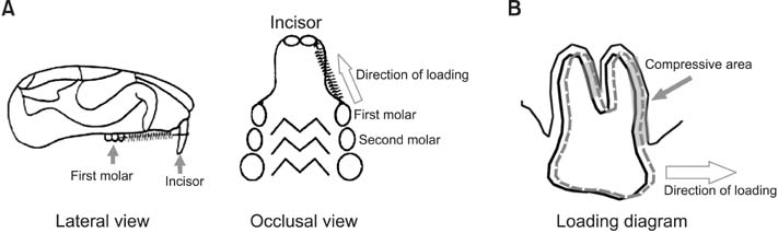
Figure 1 A rat model of orthodontic tooth movement can provide compressive force on alveolar bone. A nickel-titanium closed-coil spring exerting an orthodontic force was ligated unilaterally to the maxillary first molar and incisor. The maxillary first molar on the other side was without movement and was used as the control group. A, Lateral and occlusal view of the rat maxillae with appliance. B, The principle of orthodontic tooth movement: when the tooth was subjected to mechanical loading, some compression areas occurred on the alveolar bone surface around the tooth.
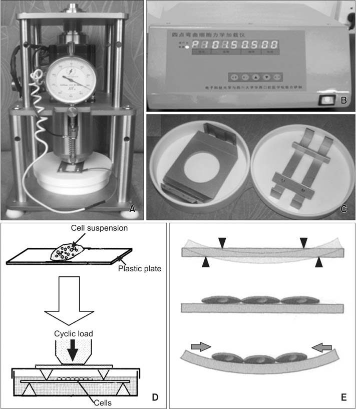
Figure 2 A four-point bending system can provide cyclic uniaxial compressive force on adherent cells in vitro. A, Actuator. B, Digital-control part. C, Culture dish with pressure head. D, Schematic representation of the apparatus used for application of mechanical force to cell cultures. The cell suspension was plated on a plastic plate, then, after 2 hours of cultivation, cells were subjected to cyclic compressive force in culture medium. E, The principle of the four-point bending system used for compressive force application (conceptual diagram).

Figure 3 Histological examination of the compression areas of periodontal tissues from the experimental group. Multinucleated osteoclasts were seen adjacent to the alveolar bone surface, with no osteoblasts observed. Hematoxylin and eosin stain, 200× magnification. AB, Alveolar bone; R, root of molar; OC, osteoclast.

Figure 4 Immunohistochemical analysis of ephrinB2 expression in periodontal tissues. A, Photomicrograph showing weak anti-ephrinB2 antibody immunolabeling of fibroblasts in periodontal tissues from the control group (without tooth movement). B, Photomicrograph showing strong anti-ephrinB2 antibody immunolabeling of osteoclasts in the compression area of periodontal tissues from the experimental group. C, Negative controls show no immunolabeling of osteoclasts. All images are at 200× magnification. AB, Alveolar bone; OC, osteoclast.
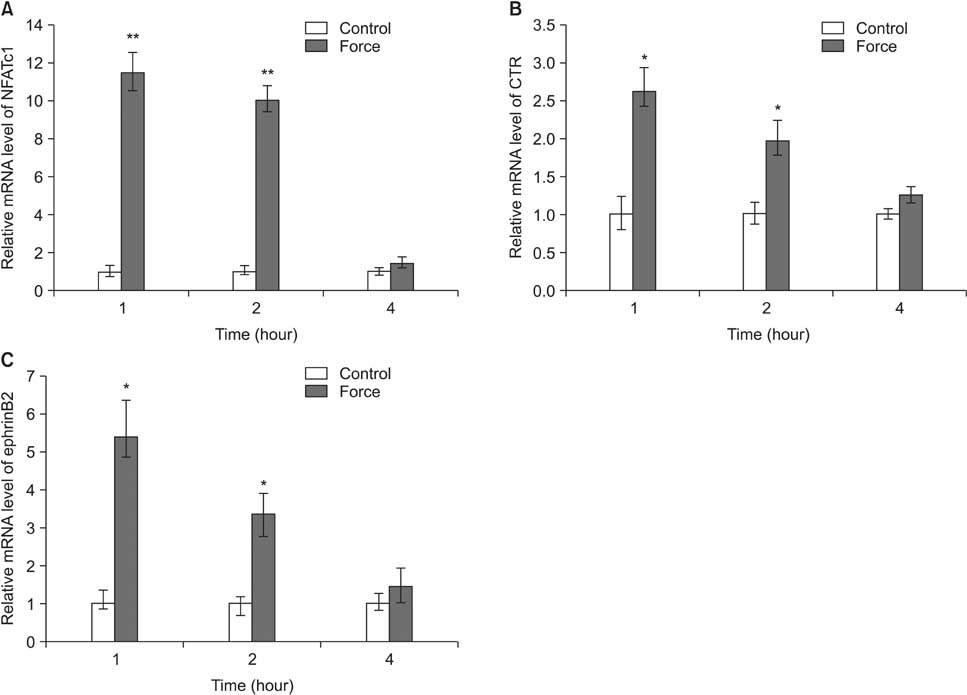
Figure 5 The effects of compressive force on osteoclastogenic gene and ephrinB2 expression in RAW264.7 cells. RAW264.7 cells stimulated with RANKL were exposed to compressive force or no force for 1, 2 and 4 hours. The mRNA expression of A, NFATc1, B, CTR, and C, ephrinB2 was then determined in control and compressed cells using quantitative real-time PCR. GAPDH was used as the internal reference gene. Data from one representative experiment of three are shown. RANKL, Receptor activator of nuclear factor kappa B ligand; NFATc1, nuclear factor of activated T cells cytoplasmic 1; CTR, calcitonin receptor; PCR, polymerase chain reaction; GAPDH, glyceraldehyde 3-phosphate dehydrogenase. *p < 0.05, **p < 0.01.
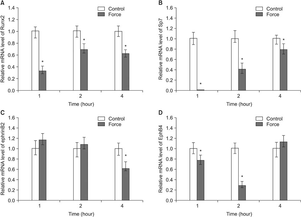
Figure 6 The effects of compressive force on the expression of osteogenic genes, ephrinB2, and EphB4 in ST2 cells. ST2 cells were exposed to compressive force or no force for 1, 2, and 4 hours. The mRNA expression of A, Runx2, B, Sp7, C, ephrinB2, and D, EphB4 was then determined in control and compressed cells. Glyceraldehyde 3-phosphate dehydrogenase (GAPDH) was used as the internal reference gene. Data from one representative experiment of three are shown. *p < 0.05.
Reference
-
1. Krishnan V, Davidovitch Z. Cellular, molecular, and tissue-level reactions to orthodontic force. Am J Orthod Dentofacial Orthop. 2006; 129:469.e1–469.e32.
Article2. Wise GE, King GJ. Mechanisms of tooth eruption and orthodontic tooth movement. J Dent Res. 2008; 87:414–434.
Article3. Masella RS, Meister M. Current concepts in the biology of orthodontic tooth movement. Am J Orthod Dentofacial Orthop. 2006; 129:458–468.
Article4. Roberts WE, Huja S, Roberts JA. Bone modeling: biomechanics, molecular mechanisms, and clinical perspectives. Sem Orthod. 2004; 10:123–161.
Article5. Oshiro T, Shiotani A, Shibasaki Y, Sasaki T. Osteoclast induction in periodontal tissue during experimental movement of incisors in osteoprotegerin-deficient mice. Anat Rec. 2002; 266:218–225.
Article6. Kanzaki H, Chiba M, Shimizu Y, Mitani H. Periodontal ligament cells under mechanical stress induce osteoclastogenesis by receptor activator of nuclear factor kappaB ligand up-regulation via prostaglandin E2 synthesis. J Bone Miner Res. 2002; 17:210–220.
Article7. Kanzaki H, Chiba M, Takahashi I, Haruyama N, Nishimura M, Mitani H. Local OPG gene transfer to periodontal tissue inhibits orthodontic tooth movement. J Dent Res. 2004; 83:920–925.
Article8. Harada S, Rodan GA. Control of osteoblast function and regulation of bone mass. Nature. 2003; 423:349–355.
Article9. Zhao C, Irie N, Takada Y, Shimoda K, Miyamoto T, Nishiwaki T, et al. Bidirectional ephrinB2-EphB4 signaling controls bone homeostasis. Cell Metab. 2006; 4:111–121.
Article10. Edwards CM, Mundy GR. Eph receptors and ephrin signaling pathways: a role in bone homeostasis. Int J Med Sci. 2008; 5:263–272.
Article11. Matsuo K. Eph and ephrin interactions in bone. Adv Exp Med Biol. 2010; 658:95–103.
Article12. Holder N, Durbin L, Cooke J. Eph receptors and ephrins are key regulators of morphogenesis. Ernst Schering Res Found Workshop. 2000; (29):123–147.
Article13. Pasquale EB. Eph receptor signalling casts a wide net on cell behaviour. Nat Rev Mol Cell Biol. 2005; 6:462–475.
Article14. Allan EH, Häusler KD, Wei T, Gooi JH, Quinn JM, Crimeen-Irwin B, et al. EphrinB2 regulation by PTH and PTHrP revealed by molecular profiling in differentiating osteoblasts. J Bone Miner Res. 2008; 23:1170–1181.
Article15. Martin TJ, Allan EH, Ho PW, Gooi JH, Quinn JM, Gillespie MT, et al. Communication between ephrinB2 and EphB4 within the osteoblast lineage. Adv Exp Med Biol. 2010; 658:51–60.
Article16. Obi S, Yamamoto K, Shimizu N, Kumagaya S, Masumura T, Sokabe T, et al. Fluid shear stress induces arterial differentiation of endothelial progenitor cells. J Appl Physiol (1985). 2009; 106:203–211.
Article17. Diercke K, Kohl A, Lux CJ, Erber R. Strain-dependent up-regulation of ephrin-B2 protein in periodontal ligament fibroblasts contributes to osteogenesis during tooth movement. J Biol Chem. 2011; 286:37651–37664.
Article18. Diercke K, Sen S, Kohl A, Lux CJ, Erber R. Compression-dependent up-regulation of ephrin-A2 in PDL fibroblasts attenuates osteogenesis. J Dent Res. 2011; 90:1108–1115.
Article19. Suzuki N, Yoshimura Y, Deyama Y, Suzuki K, Kitagawa Y. Mechanical stress directly suppresses osteoclast differentiation in RAW264.7 cells. Int J Mol Med. 2008; 21:291–296.
Article20. Liu J, Liu T, Zheng Y, Zhao Z, Liu Y, Cheng H, et al. Early responses of osteoblast-like cells to different mechanical signals through various signaling pathways. Biochem Biophys Res Commun. 2006; 348:1167–1173.
Article21. Takayanagi H, Kim S, Koga T, Nishina H, Isshiki M, Yoshida H, et al. Induction and activation of the transcription factor NFATc1 (NFAT2) integrate RANKL signaling in terminal differentiation of osteoclasts. Dev Cell. 2002; 3:889–901.
Article22. Asagiri M, Sato K, Usami T, Ochi S, Nishina H, Yoshida H, et al. Autoamplification of NFATc1 expression determines its essential role in bone homeostasis. J Exp Med. 2005; 202:1261–1269.
Article23. Takayanagi H. Mechanistic insight into osteoclast differentiation in osteoimmunology. J Mol Med (Berl). 2005; 83:170–179.
Article24. Lee SK, Goldring SR, Lorenzo JA. Expression of the calcitonin receptor in bone marrow cell cultures and in bone: a specific marker of the differentiated osteoclast that is regulated by calcitonin. Endocrinology. 1995; 136:4572–4581.
Article25. Choi JY, Pratap J, Javed A, Zaidi SK, Xing L, Balint E, et al. Subnuclear targeting of Runx/Cbfa/AML factors is essential for tissue-specific differentiation during embryonic development. Proc Natl Acad Sci U S A. 2001; 98:8650–8655.
Article26. Pratap J, Galindo M, Zaidi SK, Vradii D, Bhat BM, Robinson JA, et al. Cell growth regulatory role of Runx2 during proliferative expansion of preosteoblasts. Cancer Res. 2003; 63:5357–5362.27. Nakashima K, Zhou X, Kunkel G, Zhang Z, Deng JM, Behringer RR, et al. The novel zinc finger-containing transcription factor osterix is required for osteoblast differentiation and bone formation. Cell. 2002; 108:17–29.
Article28. Kaback LA, Soung do Y, Naik A, Smith N, Schwarz EM, O'Keefe RJ, et al. Osterix/Sp7 regulates mesenchymal stem cell mediated endochondral ossification. J Cell Physiol. 2008; 214:173–182.
Article29. Waldo CM, Rothblatt JM. Histologic response to tooth movement in the laboratory rat; procedure and preliminary observations. J Dent Res. 1954; 33:481–486.
Article30. Zaki Ae, Vanhuysen G. Histology of the periodontium following tooth movement. J Dent Res. 1963; 42:1373–1379.
Article
- Full Text Links
-
- Actions
-
Cited
- CITED
-
- Close
- Share
-
- Similar articles
-
- An electron microscopic study on the alveolar bone remodelling in pressure zones of rat molar periodontium associated with orthodontic tooth movement
- Effects of bisphosphonate and indomethacin on alveolar bone remodeling in rats
- A histological and histochemical study on the periodontal tissue reaction during experimental tooth movement in the rat
- Mechanisms of Osteoclastogenesis in Orthodontic Tooth Movement and Orthodontically Induced Tooth Root Resorption
- Effect of vitamin C deficiency on the rate of orthodontic tooth movement and alveolar bone remodeling
