Dural Arteriovenous Fistula on the Brain Stem and Upper Cervical Spinal Cord: A Case Report
- Affiliations
-
- 1Department of Physical & Rehabilitation Medicine, Research Institute of Medical Sciences, Center for Aging and Geriatrics, Regional CardioCerebrovascular Center, Chonnam National University Medical School & Hospital, Gwangju 501-757, Korea. LEE9299@hitel.
- KMID: 2267238
- DOI: http://doi.org/10.5535/arm.2011.35.5.733
Abstract
- A 53-year-old man abruptly developed headache and unconsciousness. Brain computed tomography (CT) and CT angiography showed subarachnoid hemorrhage, intraventricular hemorrhage, and multiple tortuous vascular structures on the brain stem and upper cervical spinal cord. Four-vessel angiography displayed intradural ventral arteriovenous fistula, supplied by the left vertebral and occipital arteries. Drainage was via both sigmoid sinus and cervical venous plexus. He had been treated with transarterial coil embolization of the left vertebral artery. Subsequently, he suffered from left hemiplegia and cognitive problem. Brain magnetic resonance (MR) and MR angiography performed 4 weeks later revealed multiple infarctions on the left cerebellum, left upper cervical spinal cord, and both medial thalamus, as well as occlusion of the left vertebral artery with reduction in varix size. After rehabilitative management, his muscle strength and cognitive function improved. We report a very rare case of dural arteriovenous fistula on the brain stem and upper cervical spinal cord.
Keyword
MeSH Terms
-
Angiography
Arteries
Arteriovenous Fistula
Brain
Brain Stem
Central Nervous System Vascular Malformations
Cerebellum
Colon, Sigmoid
Drainage
Headache
Hemiplegia
Hemorrhage
Humans
Infarction
Magnetic Resonance Spectroscopy
Middle Aged
Muscle Strength
Spinal Cord
Subarachnoid Hemorrhage
Thalamus
Unconsciousness
Varicose Veins
Vertebral Artery
Figure
-

Fig. 1 Brain CT and CT angiography show diffuse subarachnoid hemorrhage, intraventricular hemorrhage with hydrocephalus (A, B), and tortuous vascular structures along anterior aspect of brain stem and upper cervical spinal cord (C).
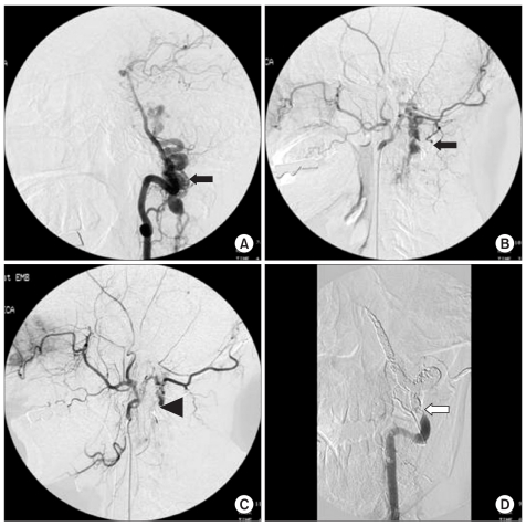
Fig. 2 Cerebral angiography shows dural arteriovenous fistula supplied by the left vertebral and occipital arteries (black arrow) (A, B). Transarterial embolization through the left occipital artery failed (black arrow head) (C). However, the left vertebral artery was super-selected with microwire and microcatheter and was embolized with embolic materials, resulting in partial occlusion (white arrow) (D).
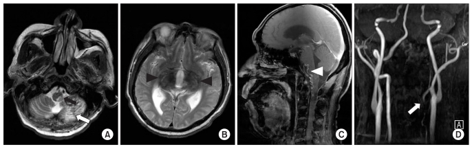
Fig. 3 Axial T2-weighted images of Brain MRI show subacute to chronic hemorrhagic infarction involving left cerebellar hemisphere (white arrow), left posterolateral aspect of upper cervical spinal cord (black arrow) (A), and both medial thalamus (black arrow head) (B). Sagittal gadolinium enhanced image shows reduction of tortuous vascular structure (white arrow head) (C). MR angiography shows occlusion of left vertebral artery (white arrow) (D).
Cited by 1 articles
-
A Rare Case of Subarachnoid Hemorrhage caused by Ruptured Venous Varix Due to Dural Arteriovenous Fistula at the Foramen Magnum Fed Solely by the Ascending Pharyngeal Artery
Hyunjun Kim, Yoon-Soo Lee, Ho-Jun Kang, Min-Seok Lee, Sang-Jun Suh, Jeong-Ho Lee, Dong-Gee Kang
J Cerebrovasc Endovasc Neurosurg. 2018;20(2):120-126. doi: 10.7461/jcen.2018.20.2.120.
Reference
-
1. Malek AM, Halbach VV, Higashida RT, Phatouros CC, Meyers PM, Dowd CF. Treatment of dural arteriovenous malformations and fistulas. Neurosurg Clin N Am. 2000; 11:147–166. PMID: 10565875.
Article2. Kan P, Stevens EA, Warner J, Couldwell WT. Resolution of an anterior-inferior cerebellar artery feeding aneurysm with the treatment of a transverse-sigmoid dural arteriovenous fistula. Skull Base. 2007; 17:205–210. PMID: 17973034.
Article3. Gilbertson JR, Miller GM, Goldman MS, Marsh WR. Spinal dural arteriovenous fistulas: MR and myelographic findings. Am J Neuroradiol. 1995; 16:2049–2057. PMID: 8585493.4. Rosenblum B, Oldfield EH, Doppman JL, Di Chiro G. Spinal arteriovenous malformation: a comparison of dural arteriovenous fistulas and intradural AVM's in 81 patients. J Neurosurg. 1987; 67:795–802. PMID: 3681418.5. Kohno M, Takahashi H, Ide K, Ishijima B, Yamada K, Nemoto S. A cervical dural arteriovenous fistula in a patient presenting with radiculopathy. J Neurosurg. 1996; 84:119–123. PMID: 8613818.
Article6. Li J, Ezura M, Takahashi A, Yoshimoto T. Intracranial dural arteriovenous fistula with venous reflux to the brainstem and spinal cord mimicking brainstem infarction-case report. Neurol Med Chir (Tokyo). 2004; 44:24–28. PMID: 14959933.
Article7. Aviv RI, Shad A, Tomlinson G, Niemann D, Teddy PJ, Molyneux AJ, Byrine JV. Cervical dural arteriovenous fistulae manifesting as subarachnoid hemorrhage: report of two cases and literature review. Am J Neuroradiol. 2004; 25:854–858. PMID: 15140735.8. Do HM, Jensen ME, Cloft HJ, Kallmes DF, Dion JE. Dural arteriovenous fistula of the cervical spine presenting with subarachnoid hemorrhage. Am J Neuroradiol. 1999; 20:348–350. PMID: 10094368.
- Full Text Links
-
- Actions
-
Cited
- CITED
-
- Close
- Share
-
- Similar articles
-
- Syringomyelia Associated with Spinal Dural Arteriovenous Fistula: Clinical and Radiological Improvement after Embolization
- Myelopathy Caused by Spinal Dural Arterio-Venous Fistula after First Lumbar Vertebral Body Fracture: A Case Report
- Dural Arteriovenous Fistula at the Foramen Magnum with Holocord Myelopathy: Case Report
- Endovascular Treatment of Spinal Dural and Epidural Arteriovenous Fistula as Complication of Lumbar Surgery
- Novalis Stereotactic Radiosurgery for Spinal Dural Arteriovenous Fistula
